

'Boiled egg' in the peritoneal cavity-a giant peritoneal loose body in a 64-year-old man: a case report
- PMID: 21736712
- PMCID: PMC3146931
- DOI: 10.1186/1752-1947-5-297
'Boiled egg' in the peritoneal cavity-a giant peritoneal loose body in a 64-year-old man: a case report
Abstract
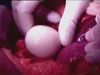
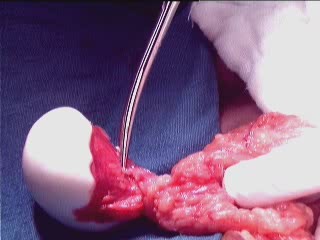
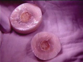
Introduction: Peritoneal loose bodies, or peritoneal mice, are rare asymptomatic lesions that are usually found as an incidental finding during abdominal surgery or autopsy. Giant loose bodies, measuring more than 5 cm, are rare and only a few cases are reported in the literature. These bodies are usually infarcted appendices epiploicae, which become detached and appear as a peritoneal loose body in the abdominal cavity. They may re-attach themselves to a surface, such as the lower aspect of the spleen or omentum, in which case they can be called a "parasitized peritoneal body", as in our case.
Case presentation: We report a case of a giant loose peritoneal body measuring 7 × 5 cm found incidentally in a 64-year-old Indian man who presented with acute intestinal obstruction. We present the current hypothesis and our opinion on the genesis of such large bodies and discuss the problems in diagnosis.
Conclusion: Peritoneal loose bodies are common but giant peritoneal loose bodies are very rare. These giant bodies usually do not require any treatment until they become complicated. Present diagnosis modalities have limitations in the diagnosis of mobile lesions in the abdominal cavity, so care must be taken to avoid unnecessary laparotomies in uncomplicated cases.
Figures
References
-
- Bhandarwar AH, Desai VV, Gajbhive RN, Deshraj BP. Acute retention of urine due to a loose peritoneal body. Br J Uro. 1996;78(6):951–952. - PubMed
LinkOut - more resources
Full Text Sources
