. 2011 Sep 29:192:413-28.
doi: 10.1016/j.neuroscience.2011.06.052. Epub 2011 Jun 26.

Central neural responses to restraint stress are altered in rats with an early life history of repeated brief maternal separation

Affiliations

Central neural responses to restraint stress are altered in rats with an early life history of repeated brief maternal separation

L Banihashemi et al. Neuroscience. .

Abstract

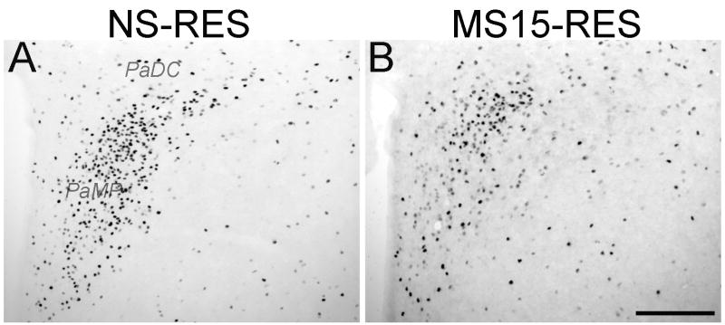
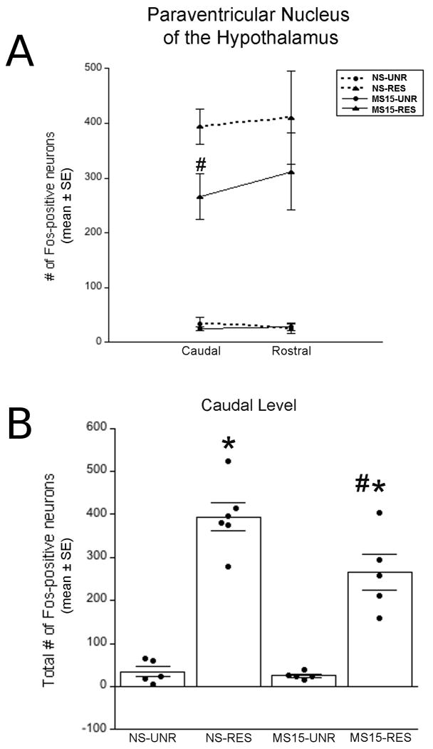
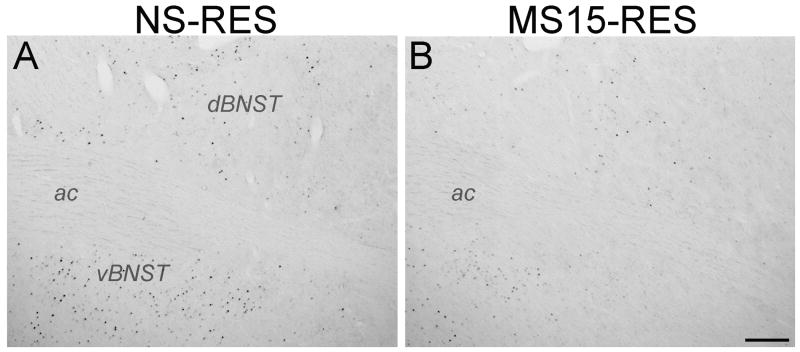
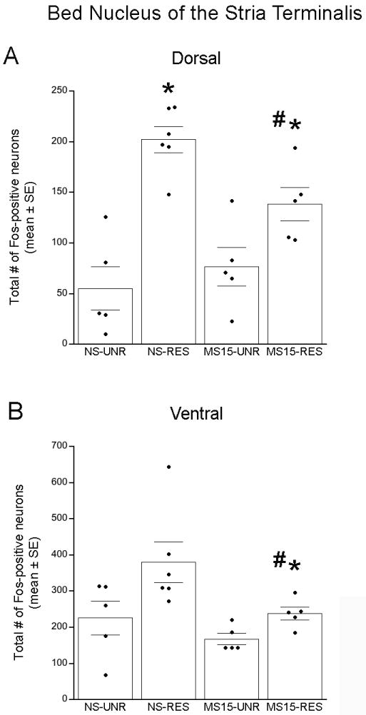
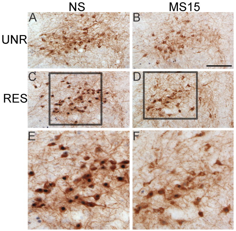
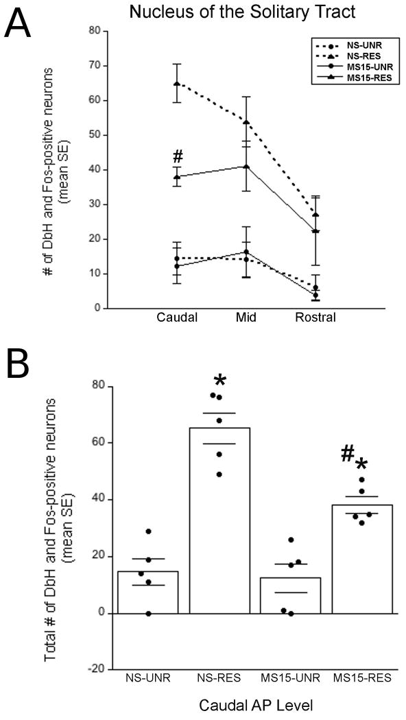
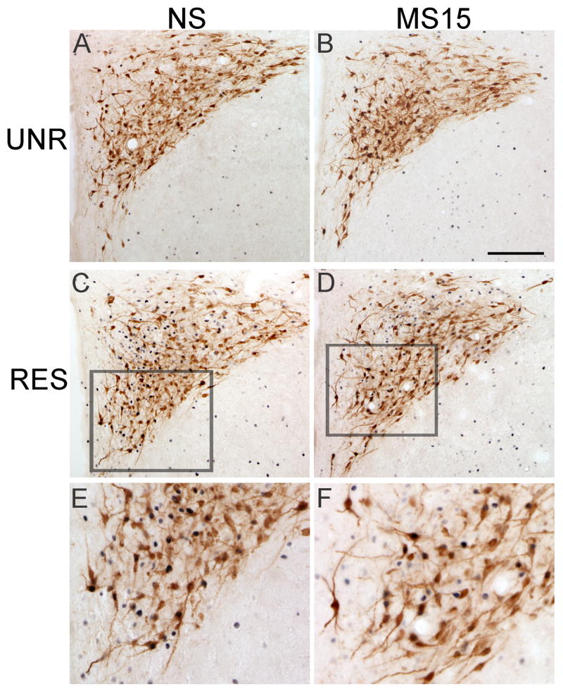
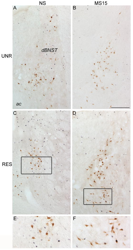
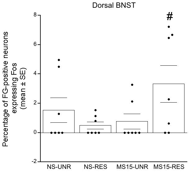
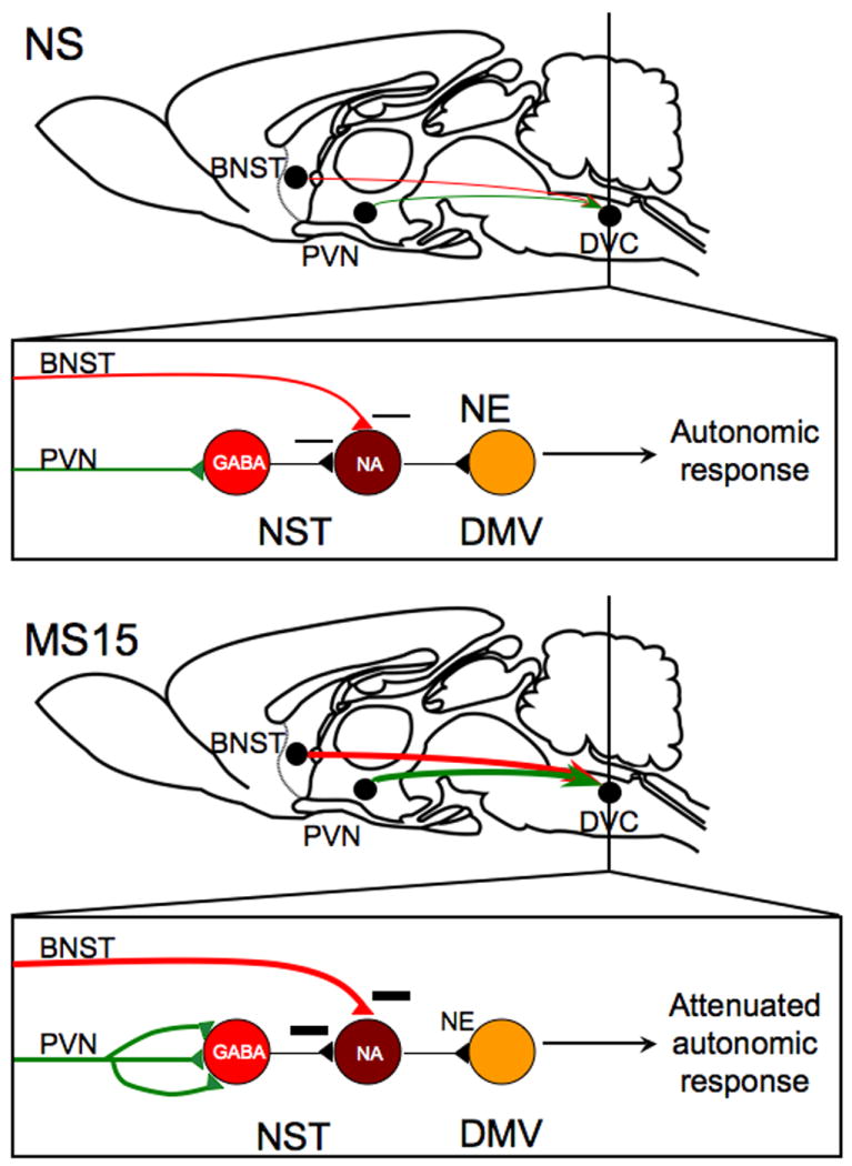
Repeated brief maternal separation (i.e. 15 min daily, MS15) of rat pups during the first one to two postnatal weeks enhances active maternal care received by the pups and attenuates their later behavioral and neuroendocrine responses to stress. In previous work, we found that MS15 also alters the developmental assembly and later structure of central neural circuits that control autonomic outflow to the viscera, suggesting that MS15 may alter central visceral circuit responses to stress. To examine this, juvenile rats with a developmental history of either MS15 or no separation (NS) received microinjection of retrograde neural tracer, FluoroGold (FG), into the hindbrain dorsal vagal complex (DVC). After 1 week, FG-injected rats and surgically intact littermates were exposed to either a 15-min restraint stress or an unrestrained control condition, and then perfused 1 h later. Brain tissue sections from surgically intact littermates were processed for Fos alone or in combination with phenotypic markers to examine stress-induced activation of neurons within the paraventricular nucleus of the hypothalamus (PVN), bed nucleus of the stria terminalis (BNST), and hindbrain DVC. Compared to NS controls, MS15 rats displayed less restraint-induced Fos activation within the dorsolateral BNST (dBNST), the caudal PVN, and noradrenergic neurons within the caudal DVC. To examine whether these differences corresponded with altered neural inputs to the DVC, sections from tracer-injected rats were double-labeled for FG and Fos to quantify retrogradely labeled neurons within hypothalamic and limbic forebrain regions of interest, and the proportion of these neurons activated after restraint. Only the dBNST displayed a significant effect of postnatal experience on restraint-induced Fos activation of DVC-projecting neurons. The distinct regional effects of MS15 on stress-induced recruitment of neurons within hypothalamic, limbic forebrain, and hindbrain regions has interesting implications for understanding how early life experience shapes the functional organization of stress-responsive circuits.


Figures

Fig. 1
Fig. 1. Photomicrographs depicting restraint-induced Fos activation within the PVN in (A) NS and (B) MS15 cases
MS15 rats displayed less restraint (RES)-induced Fos activation than NS rats. This difference was significant at the rostrocaudal level depicted in these photomicrographs (see Fig. 2). Scale bar (in B) = 200 μm, applies to both panels. PaDC; PVN dorsal cap: PaMP: PVN medial parvocellular.
Fig. 2
Fig. 2. Quantification of Fos-positive PVN neurons
(A) Line graph depicting the number of Fos-positive PVN neurons counted at two rostrocaudal levels. Compared to NS rats, MS15 rats displayed significantly less Fos activation at the caudal level of the PVN after restraint (RES). (B) Bar graph depicting the total number of Fos-positive neurons within the caudal PVN in unrestrained (UNR) control and RES cases within both postnatal groups (overlaid dot plots depict data from individual cases). RES significantly increased the number of Fos-positive PVN neurons in both postnatal groups; however, this effect was significantly attenuated in the MS15 group. # p < 0.05 between postnatal groups, * p < 0.05 within postnatal group
Fig. 3
Fig. 3. Photomicrographs depicting restraint stress-induced Fos activation within the BNST in (A) NS and (B) MS15 cases
MS15 rats displayed less restraint (RES)-induced Fos activation within the dorsolateral and ventrolateral BNST (dBNST and vBNST, respectively) compared to NS controls. Scale bar (in B) = 200 μm, applies to both panels. ac, anterior commissure.
Fig. 4
Fig. 4. Fos-positive neurons within the BNST
(A) Restraint (RES) significantly increased the number of Fos-positive neurons within the dorsolateral BNST in both postnatal groups; however, the effect of RES was significantly attenuated in the MS15 group. (B) There was a similar trend in the ventrolateral BNST, in which MS15-RS rats displayed significantly less RES-induced Fos activation compared to the NS-RES group. # p < 0.05 between postnatal groups; * p < 0.05 within postnatal group).
Fig. 5
Fig. 5. Photomicrographs depicting restraint-induced Fos activation within the NST in (A) NS and (B) MS15 cases
There was no postnatal group difference in restraint (RES)-induced Fos activation within the NST. Scale bar (in B) = 200 μm, applies to both panels. AP, area postrema; DMV, dorsal motor nucleus of the vagus; NST, nucleus of the solitary tract.
Fig. 6
Fig. 6. Photomicrographs of DbH and Fos labeling within the NST in NS (A and C) and MS15 (B and D) cases
There was no postnatal group difference in the number of double-labeled neurons among unrestrained (UNR) controls (A vs. B). However, MS15 rats displayed significantly fewer double-labeled neurons in response to restraint (RES) compared to NS controls (C vs. D). See E and F for higher-magnification views of the outlined areas in C and D. Scale bar (in B) = 200 μm, applies to panels A-D.
Fig. 7
Fig. 7. Fos and DbH-positive neurons within the NST
(A) Line graph depicting double-labeled neurons at three rostrocaudal levels of the NST. At the caudal level, there was a significant difference between the NS and MS15 groups in the number of DbH neurons activated to express Fos after restraint (RES). (B) Bar graph depicting the total number of double-labeled (DbH+Fos) neurons within the caudal level of the NST in unrestrained (UNR) control and RES rats from both postnatal groups. RES significantly increased the number of double-labeled neurons in both postnatal groups; however, this effect was attenuated in the MS15 group. Overlaid dot plots depict data from individual cases. # p < 0.05 between postnatal groups; * p< 0.05 within postnatal group.
Fig. 8
Fig. 8. Photomicrographs of FluoroGold and Fos labeling within the PVN in NS (A and C) and MS15 (B and D) cases
There was no postnatal group difference in the number of FG-positive PVN neurons projecting to the DVC (A vs. B), or in the number of FG-positive neurons expressing Fos in response to restraint (RES, C vs. D). See panels E and F for higher magnification views of the outlined areas in C and D. Scale bar (in B) = 200 μm, applies to panels A-D.
Fig. 9
Fig. 9. Photomicrographs of FluoroGold and Fos labeling within the dosolateral BNST in NS (A and C) and MS15 (B and D) cases
MS15-restrained (RES) rats displayed a greater proportion of DVC-projecting, FG-positive neurons that expressed Fos compared to NS-RES rats (C vs. D). See panels E and F for higher magnification views of the outlined areas in C and D. Scale bar (in B) = 200 μm, applies to panels A-D.
Fig. 10
Fig. 10. Bar graph depicting the percentage of DVC-projecting dosolateral BNST neurons expressing Fos
The MS15-restrained (RES) group displayed a significantly greater percentage of FG-positive neurons that expressed Fos compared to the NS-RES group. Overlaid dot plots depict data from individual cases.
Fig. 11
Fig. 11. Proposed model for MS15-related alterations in stress-related neural circuits and their potential functional consequences
Our previous finding that MS15 rats display enhanced circuit strength of PVN gastric preautonomic circuits, taken together with our current finding that MS15 rats display decreased restraint-induced activation of noradrenergic (NA) neurons within the nucleus of the solitary tract (NST), suggests that increased synaptic contacts from the paraventricular nucleus of the hypothalamus (PVN) may primarily target local GABAergic neurons within the NST. Further, our current finding that MS15 rats display increased restraint-induced activation of dorsal vagal complex (DVC)-projecting dorsolateral bed nucleus of the stria terminalis (dBNST) neurons suggests that this descending projection might contribute to decreased activation of NA NST neurons after restraint. These findings support the view that restraint stress may cause less norepinephrine (NE) release within the dorsal motor nucleus of the vagus (DMV) in MS15 rats, which may result in attenuated autonomic responses to stress, perhaps including attenuated inhibition of gastric motility.

References

    1. Abraham IM, Kovacs KJ. Postnatal handling alters the activation of stress-related neuronal circuitries. European Journal of Neuroscience. 2000;12:3003–3014. - PubMed
    1. Altschuler SM, Bao X, Bieger D, Hopkins DA, Miselis RR. Viscerotopic representation of the upper alimentary tract in the rat: sensory ganglia and nuclei of the solitary and spinal trigeminal tracts. The Journal of Comparative Neurology. 1989;283:248–268. - PubMed
    1. Aston-Jones G, Delfs JM, Druhan J, Zhu Y. The bed nucleus of the stria terminalis: a target site for noradrenergic actions in opiate withdrawal. Annals of the New York Academy of Sciences. 1999;877:486–498. - PubMed
    1. Bachelard H, Harland D, Gardiner SM, Kemp PA, Bennett T. Regional haemodynamic effects of noradrenaline injected into the hypothalamic paraventricular nuclei of conscious, unrestrained rats: possible mechanisms of action. Neuroscience. 1992;47:941. - PubMed
    1. Banihashemi L, Rinaman L. Noradrenergic inputs to the bed nucleus of the stria terminalis and paraventricular nucleus of the hypothalamus underlie hypothalamic-pituitary-adrenal axis but not hypophagic or conditioned avoidance responses to systemic yohimbine. The Journal of Neuroscience. 2006;26:11442–11453. - PMC - PubMed

Publication types