. 2011 Sep 2;286(35):30706-30713.
doi: 10.1074/jbc.M111.229831. Epub 2011 Jul 7.

DLK1-DIO3 genomic imprinted microRNA cluster at 14q32.2 defines a stemlike subtype of hepatocellular carcinoma associated with poor survival

Affiliations

DLK1-DIO3 genomic imprinted microRNA cluster at 14q32.2 defines a stemlike subtype of hepatocellular carcinoma associated with poor survival

John M Luk et al. J Biol Chem. .

Abstract

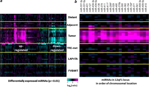
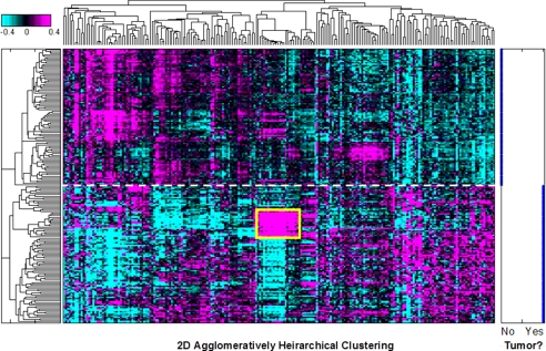
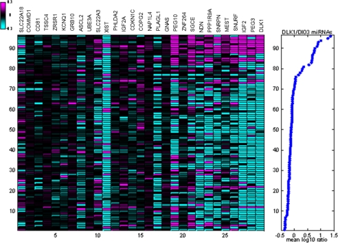
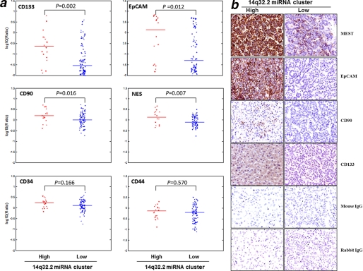
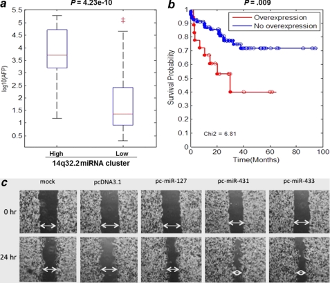
Hepatocellular carcinoma (HCC) is a heterogeneous and highly aggressive malignancy, for which there are no effective cures. Identification of a malignant stemlike subtype of HCC may offer patients with a dismal prognosis a potential targeted therapy using c-MET and Wnt pathway inhibitors. MicroRNAs (miRNAs) show promise as diagnostic and prognostic tools for cancer detection and stratification. Using a TRE-c-Met-driven transgenic HCC mouse model, we identified a cluster of 23 miRNAs that is encoded within the Dlk1-Gtl2 imprinted region on chromosome 12qF1 overexpressed in all of the isolated liver tumors. Interestingly, this region is conserved among mammalian species and maps to the human DLK1-DIO3 region on chromosome 14q32.2. We thus examined the expression of the DLK1-DIO3 miRNA cluster in a cohort of 97 hepatitis B virus-associated HCC patients and identified a subgroup (n = 18) of patients showing strong coordinate overexpression of miRNAs in this cluster but not in other cancer types (breast, lung, kidney, stomach, and colon) that were tested. Expression levels of imprinted gene transcripts from neighboring loci in this 14q32.2 region and from a subset of other imprinted sites were concomitantly elevated in human HCC. Interestingly, overexpression of the DLK1-DIO3 miRNA cluster was positively correlated with HCC stem cell markers (CD133, CD90, EpCAM, Nestin) and associated with a high level of serum α-fetoprotein, a conventional biomarker for liver cancer, and poor survival rate in HCC patients. In conclusion, our findings suggest that coordinate up-regulation of the DLK1-DIO3 miRNA cluster at 14q32.2 may define a novel molecular (stem cell-like) subtype of HCC associated with poor prognosis.


Figures

FIGURE 1.
FIGURE 1.
miRNA expression profile in the c-Met mouse model of HCC. a, tumor samples show the greatest changes in miRNA expression. miRNAs with significant expression changes in at least one sample (p < 0.01) are shown. Yellow lines separate the sample groups, and white boxes highlight the up- and down-regulated miRNAs in tumors. FVB-WT, liver tissue from control animals; LAP-tTA and TRE-met, liver tissue from single transgene parental strains; Tumor, tumor tissue from double transgene, tumor-bearing animals; Adjacent/Distant, non-tumor liver tissue from double transgene, tumor-bearing animals. b, expression pattern of 23 miRNAs from the mouse 12qF1 chromosomal cluster.
FIGURE 2.
FIGURE 2.
Global view of 220 miRNA expression patterns in tumor and non-tumor clinical tissues from human HCC. A heat map shows two-dimensional unsupervised clustering of log10 expression ratios for 220 human miRNAs in 97 pairs of tumor and adjacent non-tumor HCC tissues. miRNA expression levels were normalized to the mean of all samples. A cluster of coordinately up-regulated miRNAs from the DLK1-DIO3 region is boxed in yellow. The degree of up-regulation is shown in magenta, and down-regulation is shown in cyan on a log scale as indicated by the color bar. Separation by clustering of tumor and adjacent non-tumor samples is indicated by the dashed white line. Right, tumor status of each sample. No, adjacent non-tumor; Yes, HCC.
FIGURE 3.
FIGURE 3.
Overexpression of DLK1-DIO3 miRNA cluster at 14q32.2 is associated with up-regulation of subsets of imprinted gene transcripts. Left, log10 expression ratios in 96 HCC and matched adjacent non-tumor samples of all transcripts of genes detectably expressed in these samples and described as imprinted in humans and/or mice (17). The genes were sorted by their correlation with DLK1-DIO3 miRNA expression in matched miRNA profiles. Experiments were sorted by mean expression of DLK1-DIO3 miRNAs in matched miRNA profiles. Ratios shown are relative to mean expression in adjacent non-tumor samples. Right, the average log10 expression ratio of 22 14q32.2 miRNAs is shown for each sample. One sample pair was excluded for low quality of gene expression data.
FIGURE 4.
FIGURE 4.
DLK1-DIO3 miRNA cluster positively correlates with HCC-specific stem cell markers. a, gene expression of putative cancer stem cell markers is shown in 14q32.2 overexpression (n = 18) and no overexpression (n = 78) subgroups of HCC tumors. CD133, CD90, EpCAM, and Nestin, which have been reported as stem cell markers in HCC, were significantly correlated to the 13q32.2 miRNA expression. However, other stem cell markers, such as CD34 and CD44 (including CD29 and Nanog), did not show significant correlation. The expression levels of each marker between the 14q32.2 high and low expression groups were compared, and the p value was computed by the Wilcoxon rank sum test/Mann-Whitney U test. b, expression and localization of molecules encoded by DLK1-DIO3 imprinted microRNA cluster and stem cell markers in HCC patients. Immunohistochemistry was performed to investigate the relative expression and localization of molecules regulated by the DLK1-DIO3 imprinted microRNA cluster (MEST) in tumor tissues isolated from patients positive or negative with this microRNA cluster. In addition, the expression of stem cell markers, such as EpCAM, CD90, and CD133, was also investigated in parallel. Original magnification, ×400.
FIGURE 5.
FIGURE 5.
Overexpression of 14q32.2 miRNA cluster in HCC is associated with high serum AFP value and poor overall survival of liver cancer patients. a, analysis of variance box-and-whisker plots comparing serum AFP levels (ng/ml) (y axis) in two groups of patients. 97 HCC patients are divided into two groups by 14q32.2 microRNA expression levels; 79 show no overexpression, and 18 show overexpression. The significance of the difference in AFP level distributions is given above the plot. b, Kaplan-Meier plots comparing survival times of two groups of patients with overexpression and without overexpression of 14q32.2 miRNAs. c, in vitro wound healing assay showing that miRNAs from the DLK1-DIO3 cluster enhanced HCC cell migration potential. The PLC/PRF/5 hepatoma cell line was transfected with the selected miRNAs (miR-127, miR-431, and miR-433) as indicated or control vector. The migration of cells toward the wound was monitored, and images were captured at time 0 and 24 h. Magnification, ×100.

References

    1. Parkin D. M., Bray F., Ferlay J., Pisani P. (2005) CA Cancer J. Clin. 55, 74–108 - PubMed
    1. Lee T. K., Castilho A., Ma S., Ng I. O. (2009) Liver Int. 29, 955–965 - PubMed
    1. Sell S., Leffert H. L. (2008) J. Clin. Oncol. 26, 2800–2805 - PMC - PubMed
    1. Yang Z. F., Ho D. W., Ng M. N., Lau C. K., Yu W. C., Ngai P., Chu P. W., Lam C. T., Poon R. T., Fan S. T. (2008) Cancer Cell 13, 153–166 - PubMed
    1. Ma S., Lee T. K., Zheng B. J., Chan K. W., Guan X. Y. (2008) Oncogene 27, 1749–1758 - PubMed

MeSH terms