Review
. 2011 Sep;63(3):641-83.
doi: 10.1124/pr.110.003129. Epub 2011 Jul 7.

Activation and regulation of purinergic P2X receptor channels

Affiliations
Review

Activation and regulation of purinergic P2X receptor channels

Claudio Coddou et al. Pharmacol Rev. 2011 Sep.

Abstract

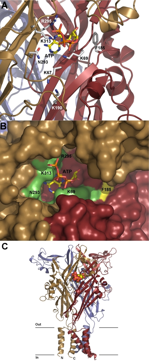
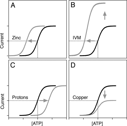
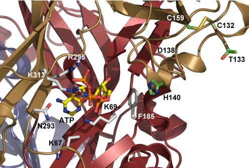
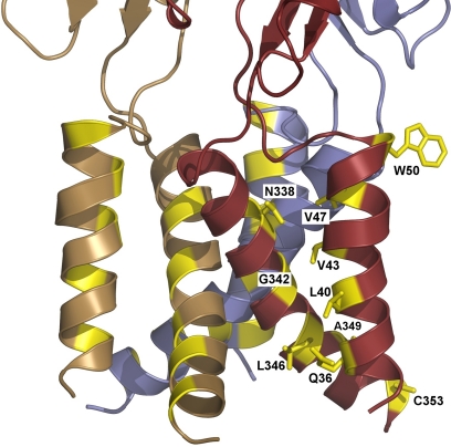
Mammalian ATP-gated nonselective cation channels (P2XRs) can be composed of seven possible subunits, denoted P2X1 to P2X7. Each subunit contains a large ectodomain, two transmembrane domains, and intracellular N and C termini. Functional P2XRs are organized as homomeric and heteromeric trimers. This review focuses on the binding sites involved in the activation (orthosteric) and regulation (allosteric) of P2XRs. The ectodomains contain three ATP binding sites, presumably located between neighboring subunits and formed by highly conserved residues. The detection and coordination of three ATP phosphate residues by positively charged amino acids are likely to play a dominant role in determining agonist potency, whereas an AsnPheArg motif may contribute to binding by coordinating the adenine ring. Nonconserved ectodomain histidines provide the binding sites for trace metals, divalent cations, and protons. The transmembrane domains account not only for the formation of the channel pore but also for the binding of ivermectin (a specific P2X4R allosteric regulator) and alcohols. The N- and C- domains provide the structures that determine the kinetics of receptor desensitization and/or pore dilation and are critical for the regulation of receptor functions by intracellular messengers, kinases, reactive oxygen species and mercury. The recent publication of the crystal structure of the zebrafish P2X4.1R in a closed state provides a major advance in the understanding of this family of receptor channels. We will discuss data obtained from numerous site-directed mutagenesis experiments accumulated during the last 15 years with reference to the crystal structure, allowing a structural interpretation of the molecular basis of orthosteric and allosteric ligand actions.


Figures

Fig. 1.
Fig. 1.
Gating properties of P2XRs. A, profiles of P2XR currents induced by sustained agonist application. Recombinant rat receptors were expressed in HEK293 cells and stimulated with ATP (10 μM for P2X1R and P2X3R, 100 μM for P2X2R and P2X4R, and 3 mM for P2X7R). τdes indicates the desensitization time constant derived from monoexponential fitting (mean ± S.E.M.; values from at least five records per channel). B to E, characterization of P2X4R current. B, Deactivation time values (10–90%) are independent from the ATP concentration. C, inverse relationship between activation time and ATP concentration. D and E, inverse relationship between the values for 10% (D) and 90% (E) desensitization time and ATP concentrations.
Fig. 2.
Fig. 2.
Dependence of the mP2X2R current on the C-terminal structure. Top, schematic representation of the P2X2R splice forms. Bottom, typical patterns of ATP-induced current profiles for P2X2Rs expressed in GT1–7 (left) and HEK293 cells (right). Blue traces, P2X2aR; red traces, P2X2bR; green traces, P2X2eR.
Fig. 3.
Fig. 3.
Characterization of rP2X3R. A, the rates of receptor desensitization. Notice the presence of the residual current during sustained agonist application. B, patterns of P2X3R current responses during repetitive stimulation with 10 nM ATP. C, time course of recovery from desensitization.
Fig. 4.
Fig. 4.
Characterization of rP2X7R. A, concentration-dependent effects of BzATP on biphasic receptor activation. B, comparison of kinetics of the secondary current growth and changes in the reversal potentials. C, sensitization of receptors induced by repetitive application of agonist. D, sensitized receptors respond with monophasic currents with the peak amplitude of current determined by agonist concentration. E, abolition of rapid and sustained P2X7R currents by the P2X7R-specific antagonist AZ10606120 (Az). F, repetitive stimulation of receptors with 100 μM BzATP in the presence and absence of AZ10606120.
Fig. 5.
Fig. 5.
Homology model of rat P2X4R with docked ATP. A, predicted structure of the ATP binding site. Residues suggested to be involved in ATP binding or located in the close vicinity of putative ATP-binding site are shown as sticks. Each subunit is shown in a different color. B, surface representation of the ATP binding pocket with a docked molecule of ATP. C, homology model of homotrimeric rat P2X4 viewed parallel to the membrane. Each subunit is depicted in a different color. The ATP molecule is shown as a Corey-Pauling-Koltun model. The black lines suggest the boundaries of the outer (out) and inner (in) layers of the plasma membrane. The homology model of rat P2X4R (sequence Arg33–Val355) was generated using the Modeler 9v7 package (Sali and Blundell, 1993) and the crystal structure of zP2X4.1R (Kawate et al., 2009) as a template. Missing side chains were built using the DeepView/Swiss-PdbViewer v4.0.1 program (Guex, 1997). Bad contacts were corrected manually and the model was energy-minimized using the DeepView/Swiss-PdbViewer with the GROMOS96 43B1 parameter set. The AutoDock v4.2 (Morris et al., 2009) was used to predict the position and the conformation of ATP within the putative ligand-binding site at the in1terface between two neighboring subunits. The figure was generated using PyMol v0.99 (http://www.pymol.org).
Fig. 6.
Fig. 6.
Patterns of rP2X4R allosteric modulations. A and C, zinc increases and protons decrease the sensitivity of receptors for agonist without affecting Emax. B, IVM treatment causes both a leftward shift in the sensitivity of receptors for agonist and an increase in the Emax value. D, copper decreases the Emax without affecting the sensitivity of receptors for agonist.
Fig. 7.
Fig. 7.
Residues of the rat P2X4R potentially involved in the modulatory effects of copper and zinc cations (Wildman et al., 1999; Acuña-Castillo et al., 2000; Coddou et al., 2003, 2007). Structural model of rat P2X4R shows the location of residues Asp138, His140, Thr133, Cys132, and Cys159 (shown as sticks) with respect to the predicted ATP-binding site. All these residues are present within the head domain of one subunit, which creates one wall of the predicted ATP-binding pocket and is probably involved in the ATP-induced conformational change. The figure was generated using PyMol v0.99 (http://www.pymol.org).
Fig. 8.
Fig. 8.
Cartoon representation of the TM domains of rat P2X4 viewed parallel to the plasma membrane plane. Residues sensitive to the presence of IVM are shown in yellow. Replacement of these residues with cysteine and alanine attenuated the allosteric action of IVM, but none of these mutations alone accounted for all of the effects of this compound on the receptor function (Jelínková et al, 2008; Jindrichova et al., 2009). For better clarity, the IVM-sensitive residues of only one subunit are shown as sticks. It is clear that the pattern of these predominantly nonpolar residues is consistent with the helical topology of both TM domains. The process of channel opening probably involves the tilting and/or rotation of TM helices (Doyle, 2004). Therefore, the mechanism of IVM action could involve facilitating the reorientation of the TM segments during the channel opening. The figure was generated using PyMol v0.99 (http://www.pymol.org).

References

    1. Acuña-Castillo C, Coddou C, Bull P, Brito J, Huidobro-Toro JP. (2007) Differential role of extracellular histidines in copper, zinc, magnesium and proton modulation of the P2X7 purinergic receptor. J Neurochem 101:17–26 - PubMed
    1. Acuña-Castillo C, Morales B, Huidobro-Toro JP. (2000) Zinc and copper modulate differentially the P2X4 receptor. J Neurochem 74:1529–1537 - PubMed
    1. Adriouch S, Bannas P, Schwarz N, Fliegert R, Guse AH, Seman M, Haag F, Koch-Nolte F. (2008) ADP-ribosylation at R125 gates the P2X7 ion channel by presenting a covalent ligand to its nucleotide binding site. FASEB J 22:861–869 - PubMed
    1. Agboh KC, Webb TE, Evans RJ, Ennion SJ. (2004) Functional characterization of a P2X receptor from Schistosoma mansoni. J Biol Chem 279:41650–41657 - PubMed
    1. Ahrens J, Leuwer M, Demir R, Krampfl K, Foadi N, Haeseler G. (2008) The anaesthetic steroid alphaxalone positively modulates alpha1-glycine receptor function. Pharmacology 82:228–232 - PubMed

Publication types

MeSH terms

LinkOut - more resources