Review
. 2011 Sep;63(3):728-49.
doi: 10.1124/pr.110.003038. Epub 2011 Jul 7.

Regulators of G-protein signaling and their Gα substrates: promises and challenges in their use as drug discovery targets

Affiliations
Review

Regulators of G-protein signaling and their Gα substrates: promises and challenges in their use as drug discovery targets

Adam J Kimple et al. Pharmacol Rev. 2011 Sep.

Abstract

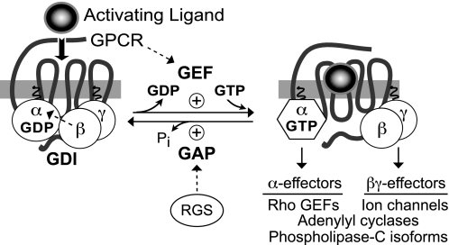
Because G-protein coupled receptors (GPCRs) continue to represent excellent targets for the discovery and development of small-molecule therapeutics, it is posited that additional protein components of the signal transduction pathways emanating from activated GPCRs themselves are attractive as drug discovery targets. This review considers the drug discovery potential of two such components: members of the "regulators of G-protein signaling" (RGS protein) superfamily, as well as their substrates, the heterotrimeric G-protein α subunits. Highlighted are recent advances, stemming from mouse knockout studies and the use of "RGS-insensitivity" and fast-hydrolysis mutations to Gα, in our understanding of how RGS proteins selectively act in (patho)physiologic conditions controlled by GPCR signaling and how they act on the nucleotide cycling of heterotrimeric G-proteins in shaping the kinetics and sensitivity of GPCR signaling. Progress is documented regarding recent activities along the path to devising screening assays and chemical probes for the RGS protein target, not only in pursuits of inhibitors of RGS domain-mediated acceleration of Gα GTP hydrolysis but also to embrace the potential of finding allosteric activators of this RGS protein action. The review concludes in considering the Gα subunit itself as a drug target, as brought to focus by recent reports of activating mutations to GNAQ and GNA11 in ocular (uveal) melanoma. We consider the likelihood of several strategies for antagonizing the function of these oncogene alleles and their gene products, including the use of RGS proteins with Gα(q) selectivity.


Figures

Fig. 1.
Fig. 1.
The standard model of guanine nucleotide cycle of G-protein coupled receptors. The heterotrimeric G-protein consists of a GDP-bound Gα subunit associated with the Gβγ heterodimer. The Gβγ serves not only to assist the coupling of Gα to the GPCR, but also as a guanine nucleotide dissociation inhibitor (GDI) for Gα, preventing the release of GDP. Upon binding of an activating ligand to the receptor, conformation changes result in the GPCR acting as a GEF, causing the release of GDP and subsequent binding of GTP. This exchange of bound nucleotide results in the dissociation of Gβγ and both Gα-GTP and Gβγ are free to signal to downstream effectors. Downstream effectors are activated until the GTP is hydrolyzed by the intrinsic GTP hydrolysis activity of the Gα subunit [which can be further accelerated by particular downstream effectors such as PLCβ1 and p115RhoGEF (Berstein et al., 1992; Kozasa et al., 1998)]. Upon hydrolysis of GTP, Gα-GDP rebinds Gβγ and the system returns to the inactive state. The rate of GTP hydrolysis can be dramatically enhanced by RGS proteins, which serve as GAPs for Gα subunits in vitro (Berman et al., 1996) and in vivo (Lambert et al., 2010).
Fig. 2.
Fig. 2.
Structural features of the heterotrimeric G-protein subunits. A, overall structural fold of the heterotrimeric G-protein Gα subunit in its inactive, GDP-bound form. The Gα subunit (PDB number 1GP2) is composed of a Ras-like domain (blue) and an all α-helical domain (green), between which is found the guanine nucleotide binding pocket (GDP in magenta). The three flexible switch regions (SI, SII, and SIII) are highlighted in cyan. B, details of structural differences between GDP- and GTP-bound states. The additional (third) phosphoryl group (orange and red) of bound GTP establishes contacts with residues Thr-181 and Gly-203 of switches I and II, respectively, thus leading to changes in all three switch regions (green; PDB number 1GIA) versus their conformation in the GDP-bound state (cyan; PDB number 1GP2). Magnesium cation is highlighted in yellow. C, overall structural fold of the Gβγ heterodimer. The Gβγ subunit (PDB number 1OMW) is colored to highlight the seven WD40 repeats that comprise the β-propeller fold: WD1, green; WD2, purple; WD3, cyan; WD4, orange; WD5, gray; WD6, wheat; and WD7; blue. The cysteine residue within Gβγ (red) that receives post-translational geranylgeranylation is highlighted in sticks configuration. The relative positioning of the N-terminal α-helix of the Gα subunit (when in the Gα-GDP/Gβγ heterotrimeric complex) is also highlighted. D, structural basis of GTP hydrolysis by Gα. Residues within Gα that are critical to the GTP hydrolysis mechanism include Arg-178 and Thr-181 from switch I and Gln-204 from switch II (colored as in A and numbered as in Gαi1; coordinates are from PDB number 1GFI). Magnesium cation is highlighted in yellow. The planar anion AlF4, which mimics the γ-phosphate leaving group of the GTP → GDP + Pi hydrolysis reaction, is depicted in metallic red.
Fig. 3.
Fig. 3.
RGS proteins stabilize the transition state of Gα subunits and coordinate the positioning of the Gα catalytic glutamate residue that is critical to intrinsic GTPase activity. Diagram of the RGS8/Gαi3 structure [PDB number 2ODE (Soundararajan et al., 2008)], as rendered using PyMOL (Schrödinger, Inc., Portland, OR). The all-helical subdomain of Gαi3 is shown in green, whereas the Ras-like nucleotide binding domain in shown in dark blue. The three flexible switch regions (SI, SII, and SIII) are highlighted in cyan. The guanine nucleotide, AlF4, and Mg2+ are highlighted in magenta, red, and yellow, respectively, whereas RGS8 is illustrated in orange. A, the RGS8 Gα-binding interface consists primarily of the SI and SII regions of Gαi3. B, the Asn-122 amide forms a hydrogen bond with Gln-204 of Gαi3, orienting it to help stabilize the planar leaving group, whereas the Asn-82 of RGS8 forms contacts with side-chain carbonyl of Thr-182, allowing the side-chain carbonyl to make a contact with the SII Lys-210 of Gαi3, stabilizing SI and SII in their transition state orientations. In addition, Asp-157 of RGS8 stabilizes the backbone amine of Thr-182, allowing the Thr-181 side-chain hydroxyl group to stabilize the Mg2+ ion (yellow).
Fig. 4.
Fig. 4.
Subfamily categorizations of the 37 RGS domain-containing proteins identified in humans, based on sequence similarities and domain architectures. An unrooted dendrogram was generated using ClustalW (http://www.clustal.org; Thompson et al., 1994) and visualized using TreeView (Page, 1996). Domain boundaries were predicted using the SMART database (Letunic et al., 2009). PDZ, PSD-95/Dlg/ZO-1 domain; PTB, phosphotyrosine-binding domain; RBD, Ras-binding domain; DEP, Dishevelled/EGL-10/Pleckstrin domain; GGL, Gγ-like domain; βCat, β-catenin interaction region; GSK3β, glycogen synthase kinase-3β interaction region; PP2A, protein phosphatase 2A; DIX, Dishevelled interaction region; SNX, sorting nexin; TM, transmembrane domain; PXA, domain associated with a PX domain; PX, p40/p47-Phox homology domain; DH, Dbl-homology domain; PH, Pleckstrin-homology domain.
Fig. 5.
Fig. 5.
Use of the RGS-insensitivity (“G>S”) and fast-hydrolysis (“G>A”) point mutants of Gα in establishing the central role of GTPase acceleration by RGS proteins in their modulatory actions on GPCR signaling kinetics and sensitivity. A cell-based system employing Gα reconstitution, along with a temporally sensitive and -reversible measure of Gαβγ activation, was established to interrogate whether “non-GAP” activities of RGS proteins (illustrated as question marks in rectangles) exist that influence the kinetics of signal onset (τonset) and receptor sensitivity (EC50 for agonist) beyond the GAP activity embodied by the RGS domain A-site. In this experimental system set up by Lambert et al. (2010), G-protein heterotrimer activation, by the binding of the agonist quinpirole to the dopamine D2 receptor (“D2-R”), increases bioluminescence resonance energy transfer (BRET) between the Gβγ-binding reporter protein masGRK3ct-Rluc8 (GRK3-ct Luc) and Gβ1γ2-Venus (YFP). Human embryonic kidney 293 cells were pretreated with pertussis toxin to inactivate native Gαi/o subunits, and so quinpirole responses were mediated by heterotrimers composed of ectopically expressed, pertussis toxin-insensitive GαoA (wt; A), PTX- and RGS-insensitive GαoA (G184S; called “G>S” in B and denoted with an asterisk), or additionally containing the fast hydrolysis switch II point mutation (G203A; called “G>A” in C). The glycine-to-alanine switch II mutation (G203A, “fast-hydrolysis”) was seen to blunt agonist sensitivity [EC50 of 1.2 μM, nearer to that of wild-type GαoA (EC50 of 405 nM)] over the more sensitive responses mediated by the use of RGS-insensitive GαoA alone (G184S; EC50 of 90 nM). In addition, normalized BRET plotted against time during sequential addition of the agonist quinpirole (30 μM) and the antagonist haloperidol (10 μM) indicated that the fast-hydrolysis mutation (“G>A”) restored rapid onset and recovery kinetics (τonset and τrecov values nearer to that of wild-type GαoA use) over the more languid responses mediated by the use of RGS-insensitive (“G>S”) GαoA alone. These results served to negate the necessity of evoking non-GAP activities of RGS proteins to explain earlier observations of RGS proteins leading to accelerated GPCR signaling onset without demonstrable changes in activating ligand potency (Doupnik et al., 1997; Saitoh et al., 1997; Zerangue and Jan, 1998), hence the removal of the question marks in rectangles from the RGS protein in the final panel.
Fig. 6.
Fig. 6.
The Gαi1 interaction surface (“A-site”) of the RGS4 RGS domain contains a charged depression. Electrostatic surface rendering of RGS4 (PDB number 1AGR; potentials contoured at ±5 kT/e), highlighting the electronegative region (red) into which the threonine-182 residue of Gαi1 is buried. Electropositive potential is highlighted in blue.
Fig. 7.
Fig. 7.
Predicted structural determinants within RGS4 that enable allosteric control over Gα-directed GAP activity by PIP3 and Ca2+/CaM. A, structural coordinates of the RGS domain of RGS4 were rendered in PyMOL using data from the RGS4/Gαi1-GDP-AlF4 complex [PDB number 1AGR (Tesmer et al., 1997a)]. The conserved RGS domain fold is composed of nine α-helices (displayed in red). Orange regions depict lysines thought to be required for PIP3 binding, whereas solid cyan areas depict the proposed A- and B-sites. Rotation about the horizontal axis by 90° is shown in the lower panel. B, electrostatic surface rendering of RGS4 (PDB number 1AGR), RGS2 (PDB number 2AF0), and RGS16 (PDB number 2BT2) using PyMol, highlighting the electropositive (blue) region considered the putative CaM-binding B-site within RGS2 and RGS4 (yellow oval). RGS16, shown to be insensitive to PIP3 and CaM modulation (Tu and Wilkie, 2004), has less electropositive potential in this region, as well as a patch of electronegative potential (red).
Fig. 8.
Fig. 8.
CCG-4986 is a generic thiol-reactive compound and should not be considered an RGS4-selective GAP-inhibitory drug. Mass spectrometry data from recombinant RGS4 (green) incubated with CCG-4986 (Kimple et al., 2007) indicates that at least two, surface-exposed cysteine residues (Cys-71 and Cys-132) become covalently modified with a 153-Da moiety (magenta) derived from the 4-nitrobenzenethiol radical liberated from CCG-4986. One of these positions (Cys-132) is near two arginines (Arg-86, Arg-90; orange) within the all α-helical domain of the Gα subunit (rendered here in a gray, space-filling translucent cloud overlying the Cα ribbon trace). Figure was rendered in PyMol based on the structural coordinates of the RGS4/Gαi1-GDP-AlF4 complex (PDB id 1AGR).
Fig. 9.
Fig. 9.
HTS-compatible enzymatic assay developed for the Gα/RGS domain target using a rate-altered Gα mutant and a fluorescence polarization immunoassay for the detection of generated GDP. The Gα subunit used in this assay bears two point mutations (denoted with asterisk) that dampen intrinsic GTPase and enhance spontaneous GDP release, respectively, thereby shifting the rate-limiting step in steady-state GTP hydrolysis away from product release [koff(GDP)] toward GTP hydrolysis [kcat(GTPase)] so that the influence of RGS domain GAP activity can be observed (section III.C.3). Fluorescent tracer is illustrated with a jagged oval; when bound to the GDP-selective monoclonal antibody, emitted light remains polarized, whereas there is low polarization of emitted light when tracer is displaced by free GDP as generated by the reaction.
Fig. 10.
Fig. 10.
Shared structural determinants of binding and GAP activity on activated Gαq, as elucidated via recent high-resolution structures of Gαq/PLCβ3 and Gαq/p63RhoGEF complexes. A, the two main effectors of Gαq-mediated signaling, PLCβ (yellow) and p63RhoGEF (salmon), share a similar helix-turn-helix configuration in their binding sites for activated Gαq and engage the same hydrophobic cleft circumscribed by the switch II (SII) (α2) and α3 helices of Gα. Structural coordinates were rendered in PyMol using PDB numbers 2RGN and 3OHM. Elements of Gαq are colored similarly to the Gα illustrations of Fig. 2. SI, switch I. B, PLCβ isoforms are not only effectors but also GAPs for Gαq/11 subunits (section I.C.4). The crystal structure of the Gαq/PLCβ3 complex revealed a mechanism for accelerating Gα GTPase activity similar to that of the RGS proteins such as RGS4 (light orange; PDB number 1AGR): namely, helping to orient the critical hydrolytic glutamine residue (Gln-209 in Gαq) via contact with an asparagine residue (Asn-260 in PLCβ3, Asn-128 in RGS4). This Gln-209 residue is mutated in Gαq and Gα11 with high frequency in uveal melanoma, so it is unlikely that the GAP activity of PLCβ or a Gαq/11-selective RGS protein would be beneficial in a gene-therapy strategy for uveal melanoma (section IV.A). Magnesium cation and water are illustrated as yellow and red spheres, respectively. C, consistent with the Gαq/PLCβ3 and Gαq/p63RhoGEF structures, the α2-α3 groove of activated Gα subunits is recognized by other effector molecules. In the GTP-bound state, switch II of Gα (α2 helix in blue) is oriented alongside the α3 helix (red), providing a hydrophobic groove that is used by diverse effector molecules (green). Left, transducin-α engages the inhibitory γ subunit of retinal phosphodiesterase (PDB number 1FQJ). Middle, Gαs recognizes adenylyl cyclase (PDB number 1AZS). Right, the phage display peptide KB1753 binds to activated Gαi1 (PDB number 2G83). These three effector molecules each insert a hydrophobic side chain into the α2-α3 groove (Trp-70, Phe-991, and Ile-9, respectively), suggesting this α2-α3 groove position as a potential site for small molecule manipulation of the Gα subunit/effector interaction.

References

    1. Anderson GR, Posokhova E, Martemyanov KA. (2009) The R7 RGS protein family: multi-subunit regulators of neuronal G protein signaling. Cell Biochem Biophys 54:33–46 - PMC - PubMed
    1. Anger T, Zhang W, Mende U. (2004) Differential contribution of GTPase activation and effector antagonism to the inhibitory effect of RGS proteins on Gq-mediated signaling in vivo. J Biol Chem 279:3906–3915 - PubMed
    1. Apanovitch DM, Slep KC, Sigler PB, Dohlman HG. (1998) Sst2 is a GTPase-activating protein for Gpa1: purification and characterization of a cognate RGS-Galpha protein pair in yeast. Biochemistry 37:4815–4822 - PubMed
    1. Arshavsky VY, Lamb TD, Pugh EN., Jr (2002) G proteins and phototransduction. Annu Rev Physiol 64:153–187 - PubMed
    1. Ashihara E, Kawata E, Maekawa T. (2010) Future prospect of RNA interference for cancer therapies. Curr Drug Targets 11:345–360 - PubMed

Publication types

MeSH terms

LinkOut - more resources