. 2011 Jul;22(7):1240-51.
doi: 10.1681/ASN.2010111149.

Podocyte COX-2 exacerbates diabetic nephropathy by increasing podocyte (pro)renin receptor expression

Affiliations

Podocyte COX-2 exacerbates diabetic nephropathy by increasing podocyte (pro)renin receptor expression

Huifang Cheng et al. J Am Soc Nephrol. 2011 Jul.

Abstract

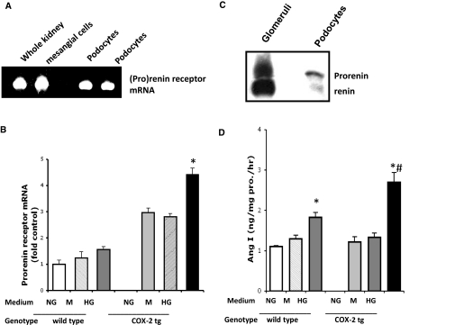
Diabetic nephropathy (DN) increases podocyte cyclooxygenase-2 (COX-2) expression, and COX-2 inhibition reduces proteinuria and glomerular injury in animal models of diabetes. To investigate the role of podocyte COX-2 in development of diabetic nephropathy, we employed a streptozotocin model of diabetic mellitus in wild-type and transgenic mice expressing COX-2 selectively in podocytes. Progressive albuminuria developed only in diabetic COX-2 transgenic mice despite hyperglycemia, BP, and GFR being similar to those in wild-type mice. Transgenic mice also manifested significant foot-process effacement, moderate mesangial expansion, and segmental thickening of the glomerular basement membrane. In cultured podocytes overexpressing COX-2, high glucose induced cell injury and increased both expression of the pro(renin) receptor and activation of the renin-angiotensin system. Downregulation of the (pro)renin receptor attenuated the injury induced by high glucose. In vivo, podocyte pro(renin) receptor expression increased in diabetic COX-2-transgenic mice, and treatment with a COX-2 inhibitor abrogated the upregulation of (pro)renin receptor and reduced albuminuria, foot-process effacement, and mesangial matrix expansion. In summary, these results demonstrate that increased expression of podocyte COX-2 predisposes to diabetic glomerular injury and that the (pro)renin receptor may be one mediator for this increased susceptibility to injury.


Figures

Figure 1.
Figure 1.
Increased COX-2 and (pro)renin receptor in db/db mice with eNOS deficiency. (A) Increased COX-2 and (pro)renin receptor expression in the kidney from db/db mice with eNOS deficiency. Immunohistochemical staining with a specific COX-2 antibody demonstrated that COX-2 is hardly detectable in normal mouse kidney (wild type in left panel); it is increased in db/db mice with eNOS deficiency (eNOS−/− db/db in right panel), not only in the macula densa (indicated by arrow head) but also at podocytes (indicated by arrows) and mesangial cells. (B) Increased (pro)renin receptor mRNA in glomeruli from eNOS−/− db/db mice. n = 4, P < 0.05. (C) Immunoreactive (pro)renin receptor cells included podocytes (indicated by arrow), endothelial cells (indicated by double head arrow), and mesangium (indicated by arrow head) within the glomeruli of eNOSKO db/db mice. KO, knockout.
Figure 2.
Figure 2.
STZ induced diabetes in wild-type and COX-2 transgenic mice. (A) STZ induced hyperglycemia in both wild-type and COX-2 transgenic mice. Wt, wild type; tg, transgenic mice with selectively increased COX-2 expression in podocytes. *P < 0.05, compared with control. There was no significant difference between Wt and COX-2 tg either at basal levels or after STZ injection. n = 5 to 6. (B) Systolic BP was not increased after 16 weeks of STZ administration. n = 5, NS. (C) GFRs were not different between groups after 16 weeks of diabetes. n = 5, NS. (D) Albuminuria was significantly increased in diabetic COX-2 tg mice. n = 6, *P < 0.05. (E) Foot process effacement was significantly increased in diabetic COX-2 tg mice. The lower right panel (HP) represents the GBM from diabetic COX-2 tg mice under higher magnification. (F) Percentage of foot process effacement. n = 3, *P < 0.05.
Figure 3.
Figure 3.
Diabetes led to upregulation of endogenous COX-2 in podocytes of COX-2 transgenic mice. (A) Immunoblotting demonstrated that COX-2 was significantly increased after STZ administration in glomeruli from COX-2 transgenic mice. n = 4, *P < 0.05, compared with wild type; #P < 0.05, compared with the transgenic control mice. (B and C) RT-PCR indicated that diabetes induced significant upregulation of endogenous COX-2 in COX-2 tg mice. n = 4, **P < 0.01 compared with wild control or basal level (B), but did not alter COX-2 transgene expression, n = 5, NS (C). (D) Increased glomerular COX-2 was primarily located in podocytes. The upper panel is representative immunohistochemical staining. COX-2 immunofluorescence is green and nephrin is red.
Figure 4.
Figure 4.
High-glucose medium induced more injury in cultured COX-2 tg podocytes. (A) High glucose induced cytoskeleton reorganization. The upper panel is from wild type and the lower from COX-2 tg podocytes. High glucose, but not mannitol, caused cytoskeleton disorganization in both wild-type and COX-2 tg podocytes but more stress fibers were seen in COX-2 tg podocytes. The lower right panel is enlarged from a single COX-2 tg podocyte exposed to high glucose. The arrows indicate cytoskeleton reorganization with loss of stress fibers and cytoplasmic aggregates. (B) High glucose decreased α-actinin 4 expression in COX-2 tg podocytes. n = 4, *P < 0.05. (C) High glucose induced more severe apoptosis in COX-2 tg podocytes. Apoptosis was determined by the percentage of TUNEL-positive cells. *P < 0.05 compared with basal level; #P < 0.05 compared with wild type in high-glucose medium. NG, normal glucose medium (5.5 mM glucose); M, 30 mM mannitol; HG, high glucose (30 mM).
Figure 5.
Figure 5.
High glucose increased (pro)renin receptor mRNA expression in COX-2 tg podocytes. (A) (Pro)renin receptor was expressed in cultured podocytes. Mouse whole kidney and primary cultured mesangial cells were used as positive controls. (B) High glucose upregulated (pro)renin receptor mRNA in COX-2 tg podocytes. n = 4, *P < 0.05. (C) Immunoreactive renin was detectable in cultured podocytes. (D) High-glucose medium stimulated renin activity, especially in COX-2 tg podocytes. n = 6, *P < 0.05 compared with basal level; #P < 0.05 COX-2 tg versus Wt.
Figure 6.
Figure 6.
COX-2 inhibition and (pro)renin receptor mRNA knockdown attenuated high-glucose–induced COX-2 tg podocyte injury. (A) High-glucose–stimulated (pro)renin receptor mRNA upregulation in COX-2 tg podocytes was significantly blunted by the COX-2 inhibitor, SC58236. n = 4, *P < 0.05 compared with wild type; #P < 0.05 compared with SC58236 treatment. (B) SC58236 also inhibited high-glucose–mediated pERK and p38 expression. Representative photograph from three separate experiments. (Pro)renin receptor mRNA knockdown partially inhibited mitogen-activated protein kinase activation (C), with scrambled SiRNA as a negative control and attenuated high-glucose–induced α-actinin 4 downregulation (D) protected from apoptosis in COX-2 tg podocytes by high glucose (E). Blue indicates DAPI staining. DAPI, 4,6-diamidino-2-phenylindole; ERK, extracellular signal–regulated kinase.
Figure 7.
Figure 7.
Diabetes stimulated (pro)renin receptor up-regulation in COX-2 tg. (A) Diabetes induced (pro)renin receptor mRNA elevation in COX-2 tg mice. n = 4, *P < 0.05 versus basal; #P < 0.05 versus Wt. (B) Diabetes led to minor increases in immunoreactive (pro)renin receptor in wild-type mice, predominantly in the mesangial area but markedly increased (pro)renin receptor in COX-2 tg mice in podocytes as well as mesangial cells. (C) Immunoflurorescence indicated colocalization of COX-2 (red) and (pro)renin (green).
Figure 8.
Figure 8.
COX-2 inhibition ameliorated diabetic renal injury in COX-2 tg mice. (A) The COX-2 inhibitor, SC58236, attenuated albuminuria in diabetic COX-2 tg mice. n = 5 to 6, *P < 0.05. (B and C) SC58236 treatment prevented diffuse foot process effacement in diabetic COX-2 tg mice. (D) SC58236 administration did not alter glomerular COX-2 expression. (E and F) SC58236 treatment inhibited glomerular (pro)renin receptor mRNA (n = 4, P < 0.05) (E) as well as immunoreactive protein staining (F). SC, SC58236.

References

    1. Tan AL, Forbes JM, Cooper ME: AGE, RAGE, and ROS in diabetic nephropathy. Semin Nephrol 27: 130–143, 2007 - PubMed
    1. Kanwar YS, Wada J, Sun L, Xie P, Wallner EI, Chen S, Chugh S, Danesh FR: Diabetic nephropathy: Mechanisms of renal disease progression. Exp Biol Med (Maywood) 233: 4–11, 2008 - PubMed
    1. Marshall SM: The podocyte: A major player in the development of diabetic nephropathy? Horm Metab Res 37 [Suppl 1]: 9–16, 2005 - PubMed
    1. Marshall SM: The podocyte: A potential therapeutic target in diabetic nephropathy? Curr Pharm Des 13: 2713–2720, 2007 - PubMed
    1. Reddy GR, Kotlyarevska K, Ransom RF, Menon RK: The podocyte and diabetes mellitus: Is the podocyte the key to the origins of diabetic nephropathy? Curr Opin Nephrol Hypertens 17: 32–36, 2008 - PubMed

Publication types

MeSH terms