. 2011 Aug 15;22(16):2912-23.
doi: 10.1091/mbc.E11-01-0017. Epub 2011 Jul 7.

Insights into EB1 structure and the role of its C-terminal domain for discriminating microtubule tips from the lattice

Affiliations

Insights into EB1 structure and the role of its C-terminal domain for discriminating microtubule tips from the lattice

Rubén M Buey et al. Mol Biol Cell. .

Abstract

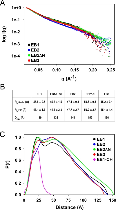
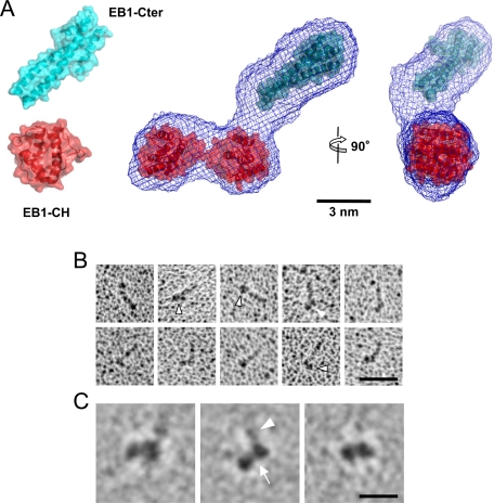
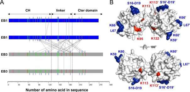
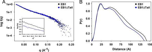
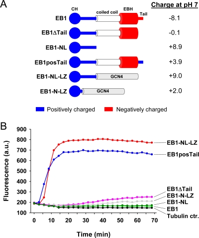
End-binding proteins (EBs) comprise a conserved family of microtubule plus end-tracking proteins. The concerted action of calponin homology (CH), linker, and C-terminal domains of EBs is important for their autonomous microtubule tip tracking, regulation of microtubule dynamics, and recruitment of numerous partners to microtubule ends. Here we report the detailed structural and biochemical analysis of mammalian EBs. Small-angle X-ray scattering, electron microscopy, and chemical cross-linking in combination with mass spectrometry indicate that EBs are elongated molecules with two interacting CH domains, an arrangement reminiscent of that seen in other microtubule- and actin-binding proteins. Removal of the negatively charged C-terminal tail did not affect the overall conformation of EBs; however, it increased the dwell times of EBs on the microtubule lattice in microtubule tip-tracking reconstitution experiments. An even more stable association with the microtubule lattice was observed when the entire negatively charged C-terminal domain of EBs was replaced by a neutral coiled-coil motif. In contrast, the interaction of EBs with growing microtubule tips was not significantly affected by these C-terminal domain mutations. Our data indicate that long-range electrostatic repulsive interactions between the C-terminus and the microtubule lattice drive the specificity of EBs for growing microtubule ends.


Figures

Figure 1:
Figure 1:
SAXS analysis of EBs. (A) Experimental SAXS scattering profiles of EB1 (black), EB2 (blue), EB2ΔN (green), and EB3 (red). (B) Rg (calculated from Guinier plot and PDFs) and Dmax values of EB1, EB2, and EB3. (C) PDFs calculated with AutoGNOM of EB1, EB2, EB2ΔN, EB3, and EB1-CH. Colors are the same as in A; EB1-CH is in pink.
Figure 2:
Figure 2:
EB model. (A) Calculated SAXS envelope (blue mesh) of EB1. The crystal structures of the CH (red ribbon; PDB ID 1PA7) and C-terminal domains (blue ribbon; PDB ID 1WU9) of EB1 are manually docked into the map. (B) Electron micrographs of rotary metal-shadowed EB1-NL-LZ-Cort specimens. The head moiety corresponding to the CH domains is highlighted by an arrowhead. Scale bar, 50 nm. (C) Averaged images of the head domain of EB1-NL-LZ-Cort specimens shown in B. Arrow and arrowhead (middle) highlight the CH and coiled-coil domains, respectively. Scale bar, 10 nm.
Figure 3:
Figure 3:
Chemical cross-linking of EBs. (A) Intermolecular map of identified cross-links between lysine residues of EB1–EB1, EB1–EB3, and EB3–EB3 dimers. The proteins are depicted as bars, and cross-linked lysines (green vertical lines) are indicated by gray lines. Monolinks (blue vertical lines) and tryptic cleavage sites (arginine; red vertical lines) are marked. (B) Model of the CH domain pair. Red and blue residues were found to be cross-linked by DSS and to interact with microtubules (Slep and Vale, 2007), respectively.
Figure 4:
Figure 4:
Role of the acidic tail for EB structure. (A) SAXS profiles and (B) PDFs of EB1 (black) and EB1ΔTail (blue). Inset in A, Guinier plots.
Figure 5:
Figure 5:
Role of the C-terminal domain in the specific enrichment of EBs at growing microtubule ends. (A) GFP-EB3 variants used in the in vitro plus end–tracking assay. The positively and negatively charged nature of the CH and C-terminal domains is indicated by blue and red, respectively. The net charge of the different GFP-tagged EB3 variants at pH 7 (based on the theoretical pI of the sequence) is indicated on the right. (B) TIRFM images and (C) kymographs of dynamic microtubules grown in the presence of the indicated concentrations of GFP-tagged EB3 protein variants. The concentration of KCl added to the buffer is also stated. Accumulation of GFP-tagged EB3-NL-LZ at the depolymerizing microtubule ends is marked by arrows. (D) Plots indicating fluorescence intensity ratios of growing microtubule ends vs. microtubule lattice for GFP-tagged EB3, EB3Δtail, and EB3-NL-LZ at different protein and salt concentrations (indicated below the plots). All measurements were performed with the same laser intensity, with the exception of 75 nM EB3-NL-LZ, for which the laser power was reduced to avoid signal saturation. (E) In vitro residence time of EB3 and its mutants on microtubule tips and lattice, measured by FRAP. All measurements were performed at 75 nM protein, with the exception of the residence time of full-length EB3 on the lattice (*), which was measured at 300 nM EB3 to obtain a sufficiently high signal to perform meaningful measurements.
Figure 6:
Figure 6:
Role of the C-terminal domain of EB1 on tubulin polymerization. (A) EB1 constructs used for the tubulin polymerization experiments. The positively and negatively charged nature of the CH and C-terminal domains is indicated by blue and red, respectively. The net charge of the different EB1 variants at pH 7 (based on the theoretical pI of the sequence) is indicated on the right. (B) Tubulin polymerization followed by DAPI fluorescence in the absence (black) and presence of EB1 (light green), EB1-NL (dark green), EB1-N-LZ (gray), EB1ΔTail (pink), EB1posTail (blue), and EB1-NL-LZ (red).

References

    1. Akhmanova A, Steinmetz MO. Tracking the ends: a dynamic protein network controls the fate of microtubule tips. Nat Rev Mol Cell Biol. 2008;9:309–322. - PubMed
    1. Akhmanova A, Steinmetz HO. Microtubule end binding: EBs sense the guanine nucleotide state. Curr Biol. 2011;21:R283–R285. - PubMed
    1. Baker NA, Sept D, Joseph S, Holst MJ, McCammon JA. Electrostatics of nanosystems: application to microtubules and the ribosome. Proc Natl Acad Sci USA. 2001;98:10037–10041. - PMC - PubMed
    1. Bieling P, Kandels-Lewis S, Telley IA, van Dijk J, Janke C, Surrey T. CLIP-170 tracks growing microtubule ends by dynamically recognizing composite EB1/tubulin-binding sites. J Cell Biol. 2008;183:1223–1233. - PMC - PubMed
    1. Bieling P, Laan L, Schek H, Munteanu EL, Sandblad L, Dogterom M, Brunner D, Surrey T. Reconstitution of a microtubule plus-end tracking system in vitro. Nature. 2007;450:1100–1105. - PubMed

Publication types

LinkOut - more resources