Review
. 2011 Jul 8;109(2):217-30.
doi: 10.1161/CIRCRESAHA.110.231225.

G protein-dependent and G protein-independent signaling pathways and their impact on cardiac function

Affiliations
Review

G protein-dependent and G protein-independent signaling pathways and their impact on cardiac function

Douglas G Tilley. Circ Res. .

Abstract

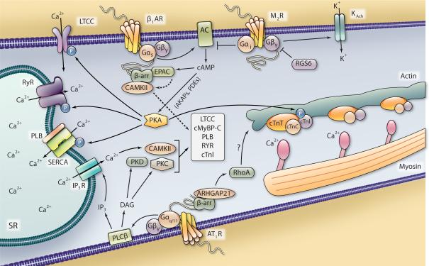
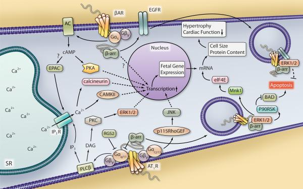
G protein-coupled receptors signal through a variety of mechanisms that impact cardiac function, including contractility and hypertrophy. G protein-dependent and G protein-independent pathways each have the capacity to initiate numerous intracellular signaling cascades to mediate these effects. G protein-dependent signaling has been studied for decades and great strides continue to be made in defining the intricate pathways and effectors regulated by G proteins and their impact on cardiac function. G protein-independent signaling is a relatively newer concept that is being explored more frequently in the cardiovascular system. Recent studies have begun to reveal how cardiac function may be regulated via G protein-independent signaling, especially with respect to the ever-expanding cohort of β-arrestin-mediated processes. This review primarily focuses on the impact of both G protein-dependent and β-arrestin-dependent signaling pathways on cardiac function, highlighting the most recent data that illustrate the comprehensive nature of these mechanisms of G protein-coupled receptor signaling.


Figures

Figure 1
Figure 1. Proposed G protein- and β-arrestin-dependent mechanisms of contractility in ventricular myocytes
Stimulation of the Gαs-coupled β1AR leads to AC-mediated generation of cAMP and increased PKA activity, which can be regulated in subcellular domains by AKAPs and PDEs. PKA signaling enhances contractility via phosphorylation of cTnI, RyR, LTCC and PLB. Modulation of the contractile machinery as well as Ca2+ entry and release of SR-stared Ca2+, which binds to the myofilaments (actin, myosin and troponin complex), act to induce contraction. A β-arrestin-dependent scaffold including EPAC and CAMKII can be recruited to β1AR upon stimulation, allowing cAMP-EPAC-mediated activation of CAMKII and regulation of contractility. Stimulation of the Gαi-coupled M2R antagonizes AC activity and releases Gβγ subunits that can open K+ channels to hyperpolarize the cardiomyocyte and dampen the contractile response, which is antagonized by RGS6. Stimulation of the Gαq/11-coupled AT1R leads to PLCβ-mediated generation of DAG, which subsequently leads to activation of PKC and PKD, and IP3, which induces the IP3R-mediated release of Ca2+ from the SR that can activate CAMKII, all of which can regulate some or all of the same myofilament and ion channel targets as PKA. β-arrestin scaffolds ARHGAP21 in response to AT1R stimulation, which leads to RhoA activation and effects on cytoskeletal structure, potentially influence cardiac contractility.
Figure 2
Figure 2. Proposed G protein- and β-arrestin-dependent regulation of ventricular myocyte hypertrophy and apoptosis
Stimulation of the AT1R leads to Gαq/11-mediated signaling that can be antagonized by RGS2 and β-arrestin recruitment. PLCβ activity leads to DAG and IP3 generation and downstream activation of PKC, ERK1/2, CAMKII and calcineurin, each of which can increase the transcriptional response in the nucleus. AT1R-Gα12/13-mediated signaling through p115RhoGEF leads to JNK activation that can also regulate transcription. βAR-Gαs stimulation leads to AC-generated cAMP accumulation and increased PKA activity, which can also modulate gene transcription. EPAC activation, possibly downstream of βAR stimulation, also leads to CAMKII and calcineurin activation via Ca2+ mobilization. Increased cardiomyocyte transcription in response to hypertrophic stimuli can lead to an increase in fetal gene expression. β-arrestin-mediated βAR signaling can also regulate hypertrophy via an unknown mechanism. Also, β-arrestin-dependent β1AR-mediated EGFR transactivation decreases cardiac apoptosis, possibly via internalization of a β1AR-EGFR-ERK1/2 complex that directs an unknown cytosolic cell survival response. Internalization of an AT1R-β-arrestin-ERK1/2 complex has been shown to increase Mnk1 activation to enhance eIF4E-mediated mRNA translation, which could contribute to an increase in cell size and protein content, thus hypertrophy and decreased cardiac function in response to hypertrophic stimuli. AT1R-β-arrestin-ERK1/2-mediated activation of p90RSK has been shown to inhibit BAD-induced apoptosis, which could contribute to cardiomyocyte cell survival.

References

    1. Penela P, Murga C, Ribas C, Tutor AS, Peregrin S, Mayor F., Jr. Mechanisms of regulation of G protein-coupled receptor kinases (GRKs) and cardiovascular disease. Cardiovasc Res. 2006;69:46–56. - PubMed
    1. Tilley DG, Rockman HA. Role of beta-adrenergic receptor signaling and desensitization in heart failure: new concepts and prospects for treatment. Expert Rev Cardiovasc Ther. 2006;4:417–432. - PubMed
    1. Ma TK, Kam KK, Yan BP, Lam YY. Renin-angiotensin-aldosterone system blockade for cardiovascular diseases: current status. Br J Pharmacol. 2010;160:1273–1292. - PMC - PubMed
    1. Kim IM, Tilley DG, Chen J, Salazar NC, Whalen EJ, Violin JD, Rockman HA. Beta-blockers alprenolol and carvedilol stimulate beta-arrestin-mediated EGFR transactivation. Proc Natl Acad Sci U S A. 2008;105:14555–14560. - PMC - PubMed
    1. Rajagopal K, Whalen EJ, Violin JD, Stiber JA, Rosenberg PB, Premont RT, Coffman TM, Rockman HA, Lefkowitz RJ. Beta-arrestin2-mediated inotropic effects of the angiotensin II type 1A receptor in isolated cardiac myocytes. Proc Natl Acad Sci U S A. 2006;103:16284–16289. - PMC - PubMed

Publication types