Review
. 2011 Jul 7;17(7):796-808.
doi: 10.1038/nm.2399.

The immunology of stroke: from mechanisms to translation

Affiliations
Review

The immunology of stroke: from mechanisms to translation

Costantino Iadecola et al. Nat Med. .

Abstract

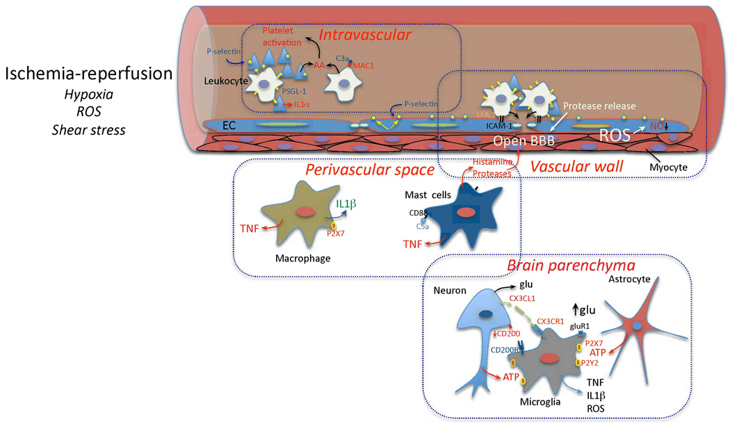
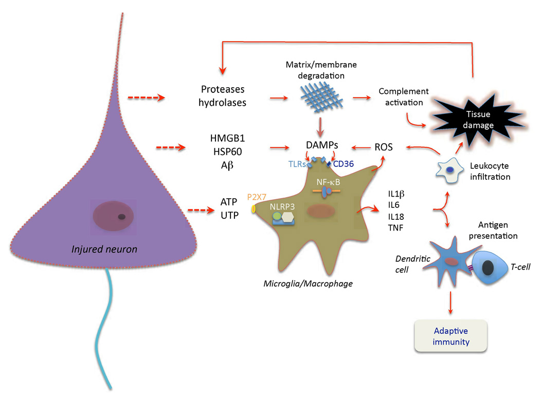
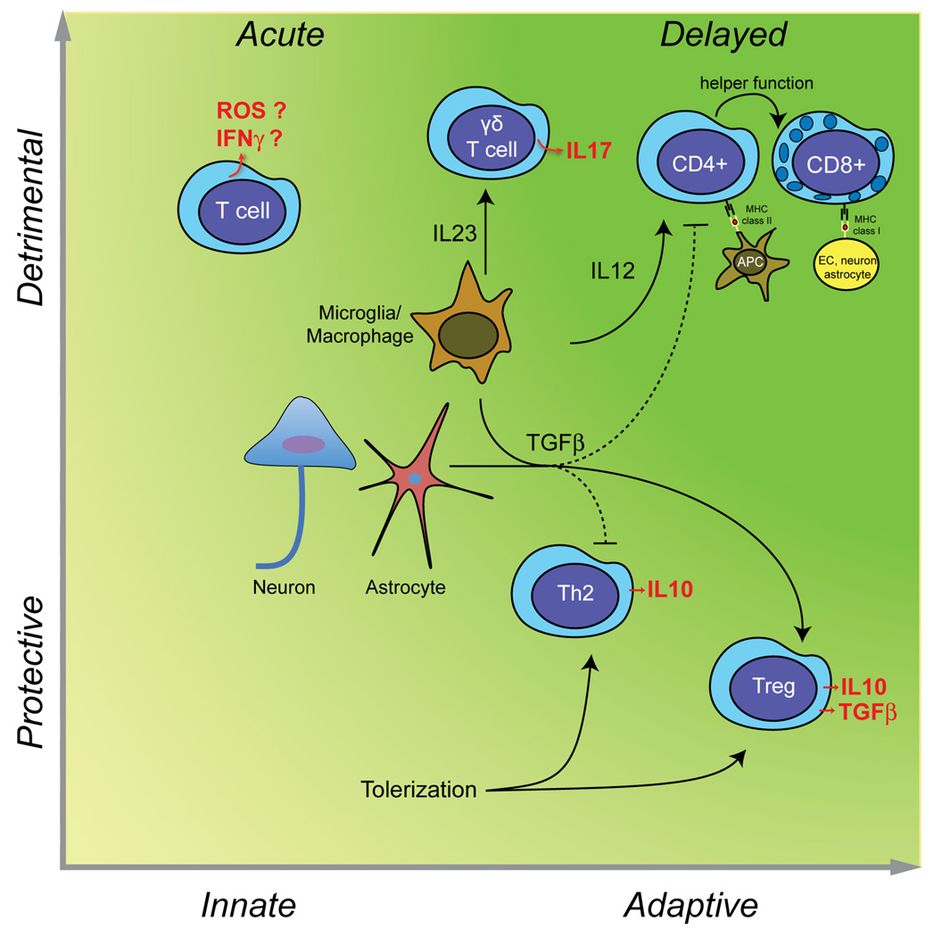
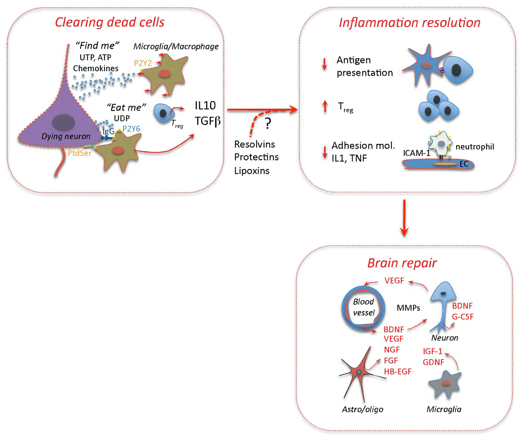
Immunity and inflammation are key elements of the pathobiology of stroke, a devastating illness second only to cardiac ischemia as a cause of death worldwide. The immune system participates in the brain damage produced by ischemia, and the damaged brain, in turn, exerts an immunosuppressive effect that promotes fatal infections that threaten the survival of people after stroke. Inflammatory signaling is involved in all stages of the ischemic cascade, from the early damaging events triggered by arterial occlusion to the late regenerative processes underlying post-ischemic tissue repair. Recent developments have revealed that stroke engages both innate and adaptive immunity. But adaptive immunity triggered by newly exposed brain antigens does not have an impact on the acute phase of the damage. Nevertheless, modulation of adaptive immunity exerts a remarkable protective effect on the ischemic brain and offers the prospect of new stroke therapies. As immunomodulation is not devoid of deleterious side effects, a better understanding of the reciprocal interaction between the immune system and the ischemic brain is essential to harness the full therapeutic potential of the immunology of stroke.


Figures

Figure 1
Figure 1. Early vascular, perivascular and parenchymal events triggered by I/R
Hypoxia, ROS and changes in shear stress initiate the cellular events induced by I/R. In the vessels lumen, I/R leads to blood clotting, platelet aggregation and cytokine release (IL-1α). Translocation of P-selectin on the surface of platelets and EC leads to platelet-leukocyte aggregation. Complement is activated and arachidonic acid metabolites are released. In the vascular wall, upregulation of E- and P-selectin on EC provides a platform for low affinity leukocyte binding through interaction with sialyl Lex moieties of glycoproteins expressed on leukocytes, e.g. PSGL-1. Firm adhesion is obtained after endothelial expression of ICAM-1 interacting with leukocyte β2 integrins (LFA-1 and Mac-1). Loss of NO promotes vasoconstriction and enhances leukocyte and platelet aggregation. MMP activation could lead to BBB breakdown and matrix proteolysis facilitating leukocyte extravasation. In the perivascular space, chemotactic complement subunits (C5a) acting on mast cell complement receptors (CD88) leads to degranulation and release of histamine and proteases, contributing to BBB leakiness. Cytokines (TNF, IL-1β) are produced by mast cells and perivascular macrophages, providing further signals to guide leukocyte migration across the vessel wall,. In the brain parenchyma, injured cells release purines (ATP), which act as early proinflammatory signals leading to production of cytokines and chemokines. Disruption of neuronal-microglial interaction (CX3CL1, CD200) and increases in extracellular glutamate (glu) acting on microglial GluR1 metabotropic receptor also contribute to the pro-inflammatory milieu.
Figure 2
Figure 2. Cell death and activation of pattern recognition receptors set the stage adaptive immunity
Release of nucleotides (ATP, UTP) from injured cells, including neurons, activates purinergic receptors on microglia and macrophages, and leads to production of pro-inflammatory cytokines. While most of these cytokines are transcriptionally induced, IL-1β and IL-18 are processed from their pro-peptides by the activity of interleukin-1 converting enzyme (ICE; caspase 1). ICE is embedded in a multi-protein complex (NLRP3 or inflammasome) and is activated by microglial P2×7 receptors. Ischemic cell death leads to the formation of danger associated molecular patterns (DAMPs) molecules, which activate TLRs, especially TLR2 and 4. DAMPs released by ischemia include HMGB1, an intracellular DNA binding protein released after cellular injury, HSP60, and β-amyloid (Aβ), among others. TLRs, in concert with scavenger receptors such as CD36, upregulate pro-inflammatory gene expression through the transcription factor NF-κB,. DAMPs also derive from matrix breakdown by lytic enzymes released from dead cells and by the action of reactive oxygen species (ROS) on lipids. The cytokine production and complement activation resulting from these events leads to increased leukocyte infiltration and enhances tissue damage, which, in turn, produces more DAMPs. Antigens unveiled by tissue damage are presented to T cells, setting the stage for adaptive immunity.
Figure 3
Figure 3. Deleterious and beneficial roles of T cells in stroke
In the acute phase of cerebral ischemia, unprimed T cells contribute to tissue damage in an antigen independent manner (innate immunity), possibly through IFNγ and ROS (left upper quadrant). γδT cells, activated by IL-23 released from microglia/macrophages, produce the cytotoxic cytokine IL-17 and contributes to acute ischemic brain injury. However, T cells can also be protective. TGFβ produced by neurons, glia, or macroglia/macrophages promotes the development of Treg cells secreting the protective cytokine IL-10 and inhibits Th1 and Th2 responses. Treg cells are protective in models of cerebral ischemia. Induction of mucosal tolerance with CNS antigens produces an adaptive response, which leads to the establishment of autoreactive Th2 cells producing IL-10 and Treg cells producing IL-10 and TGFβ, is highly protective in experimental stroke (right lower quadrant). As discussed in the text, there is no evidence that adaptive immunity contributes to acute ischemic brain injury. However, weeks and months after stroke, autoreactive CD4+ and CD8+ T cells targeting CNS antigens could develop (right upper quadrant). The resulting cell death could play a role in the delayed brain damage and atrophy that occurs after stroke.
Figure 4
Figure 4. Resolution of inflammation and tissue repair
Clearing of dead cells and suppression of inflammation are key events in brain repair. “Find-me signals” (UTP, ATP) attract microglia and macrophages through P2Y2 receptors. “Eat-me signals” include UDP, which act on P2Y6 receptors to stimulate microglial phagocytosis, and phosphatidylserine (PtdSer), which is translocated to the outer leaflet of the plasma membrane of apoptotic cells. PtdSer binding proteins involved in the clearance of dead cells include MGF-E8 on microglia and TIM4 on macrophages. Immunoglobulins directed against CNS antigens, which appear after stroke (Table 2), may also promote phagocytosis by engaging Fc receptors on phagocytic cells. Phagocytosis promotes secretion of IL-10 and TGFβ, which, in turn, suppress antigen presentation, promote Treg formation, inhibit expression of adhesion molecules in EC and production of proinflammatory cytokines,. TGFβ and IL-10 are also neuroprotective, and may facilitate brain repair processes. In addition, arachidonic and omega-3 fatty acids metabolites lipoxins, resolvins, and protectins, which play an active role in the resolution of inflammation in other organs, could also contribute to suppress post-ischemic inflammation. Growth factors and MMPs produced by EC, neurons, astrocytes, oligodendrocytes and microglia are critical molecules driving tissue reorganization and repair,.

Comment in

  • Mannose-binding lectin-the forgotten molecule?
    Osthoff M, Trendelenburg G, Eisen DP, Trendelenburg M. Osthoff M, et al. Nat Med. 2011 Dec 6;17(12):1547-8; author reply 1548. doi: 10.1038/nm.2588. Nat Med. 2011. PMID: 22146452 No abstract available.

References

    1. Mena H, Cadavid D, Rushing EJ. Human cerebral infarct: a proposed histopathologic classification based on 137 cases. Acta Neuropathol. 2004;108:524–530. - PubMed
    1. Moskowitz MA, Lo EH, Iadecola C. The science of stroke: mechanisms in search of treatments. Neuron. 2010;67:181–198. - PMC - PubMed
    1. Meisel C, Schwab J, Prass K, Meisel A, Dirnagl U. Central nervous system injury-induced immune deficiency syndrome. Nat Rev Neurosci. 2005;6:775–786. - PubMed
    1. Urra X, Cervera A, Villamor N, Planas AM, Chamorro A. Harms and benefits of lymphocyte subpopulations in patients with acute stroke. Neuroscience. 2009;158:1174–1183. - PubMed
    1. Weiner HL. The challenge of multiple sclerosis: how do we cure a chronic heterogeneous disease? Ann Neurol. 2009;65:239–248. - PubMed

Publication types

Substances