. 2011:17:1669-78.
Epub 2011 Jun 22.

A new VCAN/versican splice acceptor site mutation in a French Wagner family associated with vascular and inflammatory ocular features

Affiliations

A new VCAN/versican splice acceptor site mutation in a French Wagner family associated with vascular and inflammatory ocular features

Antoine P Brézin et al. Mol Vis. 2011.

Abstract

Purpose: To detail the highly variable ocular phenotypes of a French family affected with an autosomal dominantly inherited vitreoretinopathy and to identify the disease gene.

Methods: Sixteen family members with ten affected individuals underwent detailed ophthalmic evaluation. Genetic linkage analysis and gene screening were undertaken for genes known to be involved in degenerative and exudative vitreoretinopathies. Qualitative reverse transcriptase-PCR analysis of the versiscan (VCAN) transcripts was performed after mutation detection in the VCAN gene.

Results: The first index patient of this French family was referred to us because of a chronic uveitis since infancy; this uveitis was associated with exudative retinal detachment in the context of a severe uncharacterized familial vitreoretinopathy. Genetic linkage was obtained to the VCAN locus, and we further identified a new pathogenic mutation at the highly conserved splice acceptor site in intron 7 of the VCAN gene (c.4004-2A>T), which produced aberrantly spliced VCAN transcripts.

Conclusions: Extensive molecular investigation allowed us to classify this familial vitreoretinopathy as Wagner syndrome. This study illustrates the need to confirm clinical diagnosis by molecular genetic testing and adds new ocular phenotypes to the Wagner syndrome, such as vascular and inflammatory features.


Figures
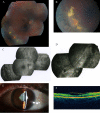
Figure 1
Figure 1
Patient V.5 (the first index case of the family). A: Mosaic multifield fundus photograph of the patient’s right eye, showing an avascular vitreous veil, approximately 2–3 disc diameters anterior to the inferior temporal retinal vein, with patches of chorioretinal atrophy, retinal pigmentary, and exudative lesions. B: Exudative yellow lesions with telangiectatic Coats-like neovascularization are seen in the retinal periphery. C: Fluorescein angiogram of the right eye of patient V.5 showing the superior pole and superior nasal and inferior temporal periphery. A mildly titled disc is seen. The retinal vasculature appears normal at the posterior pole and in the superior nasal periphery, and pigment clumping is present in the inferior temporal field. D: Fluorescein angiogram of the right eye of patient V.5 showing pigment clumping parallel to the ora in the inferior nasal periphery. Peripheral vascular leakage is seen as diffuse areas of hyperfluorescence. E: Slit-lamp photograph of the left eye of patient V.5 showing iris–lens synechiae from the 7:00 to the 10:00 position. F: OCT imaging of the right eye of patient V.5 showing a dense posterior hyaloid forming a bridge over the foveal pit.
Figure 2
Figure 2
Mosaic multifield fundus photographs of different family members. A: Fundus photograph of patient V.2 showing a nasal dragging of the retinal vessels and fovea with white perivascular sheathing. Clumps of pigment are visible in the nasal retina. No avascular vitreous membrane was seen. B: Fundus photograph of the right eye of patient V.6 showing a normal retinal vasculature with atrophy and pigment clumping along and anterior to the temporal arcades. No avascular vitreous membrane was observed. C: Fundus photograph of the left eye of patient V.6 showing diffuse pigmentation with chorioretinal atrophy.
Figure 3
Figure 3
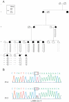
Pedigree of the Wagner family, linkage and mutation analysis of the versican (VCAN) gene. A: The pedigree of the family studied consisted in ten affected and six unaffected participating individuals, providing strong evidence of autosomal dominant mode of inheritance. Filled symbols represent affected individuals, whereas clear symbols refer to unaffected individuals. The arrow indicates the first proband examined in our ophthalmologic center. Segregation analysis of microsatellite markers encompassing the WGN1 locus on chromosome 5q14.3. The disease-associated haplotype is boxed and is shared between all affected members. The genetic markers and the corresponding allele are listed in map order. B: Partial sequence chromatograms of intron 7–exon 8 boundary of the VCAN gene from one affected individual (V.5) and one normal control subject (V.4) showing a heterozygous adenine to thymine transversion at the second base of the 3′ acceptor splice site of intron 7 (c.4004–2A>T).
Figure 4
Figure 4
Versican (VCAN) transcript analysis. A: Ethidium bromide-stained agarose gel of reverse transcription- polymerase chain reaction (RT–PCR) products obtained with primer pairs 7F/8R for amplification of the versican V0 mRNA from cultured lymphoblasts of one affected individual (V.5) and one control unaffected subject (V.4). The results indicate that both a wild-type (band of 372 bp) and a 39-nt deleted (band of 333 bp) cDNA fragment of the V0 transcript are present in the affected patient harboring the intron 7 splice acceptor mutation of the VCAN gene (c.4004–2A>T). The nucleotide change at the 3′ acceptor splice site of intron 7 of the VCAN gene was analyzed using the NNSplice program for prediction in alteration of splicing junctions, and the splice-site scores (SSSs) for a normal site (SSS-nor), a mutated site (SSS-mut), and a cryptic site (SSS-crypt) were calculated [28]. B: Partial sequence chromatogram of this aberrant V0 transcript lacking the first 39 bp of VCAN exon 8.

References

    1. Graemiger RA, Niemeyer G, Schneeberger SA, Messmer EP. Wagner vitreoretinal degeneration. Follow-up of the original pedigree. Ophthalmology. 1995;102:1830–9. - PubMed
    1. Wagner H. Ein bisher unbekanntes Erbleiden des Auges (degeneratiohyaloideo-retinalis hereditaria), beobachtet im Kanton Zurich. Klin Mbl Augenheilk. 1938;100:840–57.
    1. Stickler GB, Belau PG, Farrell FJ, Jones JD, Pugh DG, Steinberg AG, Ward LE. Hereditary progressive arthro-ophthalmopathy. Mayo Clin Proc. 1965;40:433–55. - PubMed
    1. Black GC, Perveen R, Wiszniewski W, Dodd CL, Donnai D, McLeod D. A novel hereditary developmental vitreoretinopathy with multiple ocular abnormalities localizing to a 5-cM region of chromosome 5q13-q14. Ophthalmology. 1999;106:2074–81. - PubMed
    1. Brown DM, Graemiger RA, Hergersberg M, Schinzel A, Messmer EP, Niemeyer G, Schneeberger SA, Streb LM, Taylor CM, Kimura AE. Genetic linkage of Wagner disease and erosive vitreoretinopathy to chromosome 5q13–14. Arch Ophthalmol. 1995;113:671–5. - PubMed

Publication types

MeSH terms

Supplementary concepts

LinkOut - more resources