

Expression of cell proliferation and apoptosis biomarkers in pterygia and normal conjunctiva
- PMID: 21738398
- PMCID: PMC3130721
Expression of cell proliferation and apoptosis biomarkers in pterygia and normal conjunctiva
Abstract
Purpose: To analyze the expression of apoptosis and cell proliferation molecules in pterygium tissues of Chinese patients.
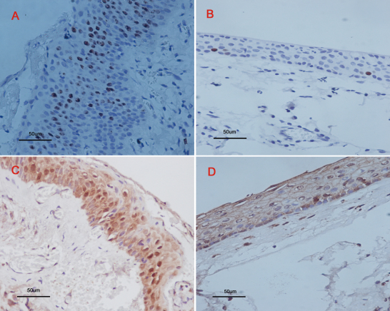
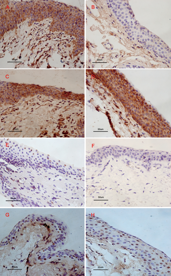
Methods: Thirty-three pterygia were surgically removed using the bare sclera procedure, and 23 normal bulbar conjunctivas were also obtained. Formalin-fixed, paraffin-wax-embedded tissues were analyzed by immunohistochemistry with anti- proliferating cell nuclear antigen (PCNA), K(i)-67 (a proliferating cell marker), mutant p53 (mP53), Bcl-2 associated X-protein (BAX), B-cell lymphoma gene 2 (Bcl-2), and caspase-3 antibodies. Terminal deoxynucleotidyl transferase-mediated dUTP-biotin nick end labeling assay (TUNEL) analysis was used to analyze the apoptotic cells.
Results: Our study revealed that the positive rate of PCNA and K(i)-67 significantly increased in the pterygium samples compared to the normal conjunctiva samples. In the molecules involved in apoptosis, the results showed that the positive rate of Bcl-2 and mP53 significantly increased in the pterygium samples. However, no difference was found between the pterygium and normal conjunctiva samples in the expression of Bax and caspase-3. Through TUNEL analysis, apoptotic cells were seen in the entire width of the epithelial layer in normal conjunctivas but were found mainly confined to the outer layer of the epithelial cells in pterygia.
Conclusions: The finding of high levels of cellular proliferation and low levels of cellular apoptosis in pterygia confirmed that both cell apoptosis and proliferation are known to play an important role in human pterygium pathogenesis.
Figures
References
-
- Di Girolamo N, Chui J, Coroneo MT, Wakefield D. Pathogenesis of pterygia: role of cytokines, growth factors, and matrix metalloproteinases. Prog Retin Eye Res. 2004;23:195–228. - PubMed
-
- Sekundo W, Droutsas K, Cursiefen C. Operative techniques for surgical treatment of primary and recurrent pterygia. Ophthalmologe. 2010;107:525–8. - PubMed
-
- Golu T, Mogoanta L, Streba CT, Pirici DN, Malaescu D, Mateescu GO, Mutiu G. Pterygium: histological and immunohistochemical aspects. Rom J Morphol Embryol. 2011;52:153–8. - PubMed
-
- Kase S, Osaki M, Jin XH, Ohgami K, Yoshida K, Saito W, Takahashi S, Nakanishi K, Ito H, Ohno S. Increased expression of erythropoietin receptor in human pterygial tissues. Int J Mol Med. 2007;20:699–702. - PubMed
-
- Kaimbo K. Surgical treatment of pterygium: 24 cases of excision. J Fr Ophtalmol. 1988;11:335–8. - PubMed
Publication types
MeSH terms
Substances
LinkOut - more resources
Full Text Sources
Research Materials
Miscellaneous