. 2011 Jun;7(6):e1002103.
doi: 10.1371/journal.ppat.1002103. Epub 2011 Jun 30.

The binding of triclosan to SmeT, the repressor of the multidrug efflux pump SmeDEF, induces antibiotic resistance in Stenotrophomonas maltophilia

Affiliations

The binding of triclosan to SmeT, the repressor of the multidrug efflux pump SmeDEF, induces antibiotic resistance in Stenotrophomonas maltophilia

Alvaro Hernández et al. PLoS Pathog. 2011 Jun.

Abstract

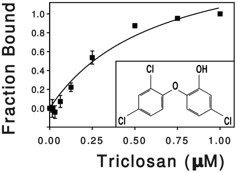
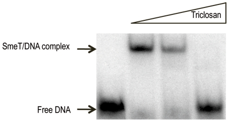
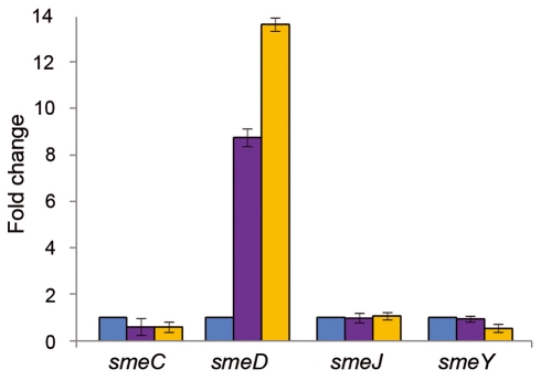
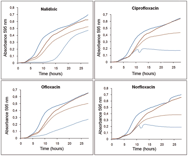
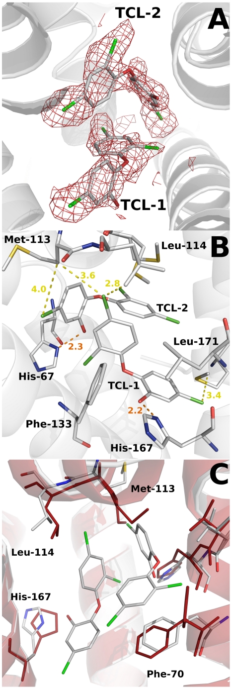
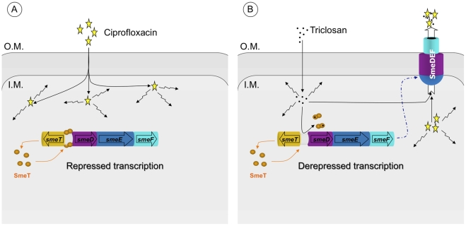
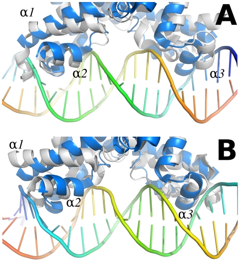
The wide utilization of biocides poses a concern on the impact of these compounds on natural bacterial populations. Furthermore, it has been demonstrated that biocides can select, at least in laboratory experiments, antibiotic resistant bacteria. This situation has raised concerns, not just on scientists and clinicians, but also on regulatory agencies, which are demanding studies on the impact that the utilization of biocides may have on the development on resistance and consequently on the treatment of infectious diseases and on human health. In the present article, we explored the possibility that the widely used biocide triclosan might induce antibiotic resistance using as a model the opportunistic pathogen Stenotrophomonas maltophilia. Biochemical, functional and structural studies were performed, focusing on SmeDEF, the most relevant antibiotic- and triclosan-removing multidrug efflux pump of S. maltophilia. Expression of smeDEF is regulated by the repressor SmeT. Triclosan released SmeT from its operator and induces the expression of smeDEF, thus reducing the susceptibility of S. maltophilia to antibiotics in the presence of the biocide. The structure of SmeT bound to triclosan is described. Two molecules of triclosan were found to bind to one subunit of the SmeT homodimer. The binding of the biocide stabilizes the N terminal domain of both subunits in a conformation unable to bind DNA. To our knowledge this is the first crystal structure obtained for a transcriptional regulator bound to triclosan. This work provides the molecular basis for understanding the mechanisms allowing the induction of phenotypic resistance to antibiotics by triclosan.


Conflict of interest statement

The authors have declared that no competing interests exist.

Figures

Figure 1
Figure 1. Effect of triclosan concentration on the fluorescence of SmeT.
SmeT was treated with increasing concentrations of triclosan at room temperature. The change in fluorescence, relative to the untreated sample, was plotted against triclosan concentration. The hyperbola shows the best fit of the data. The chemical formula of triclosan (5-chloro-2-(2,4-dichlorophenoxy) phenol) is shown into the box.
Figure 2
Figure 2. Triclosan breaks the SmeT-DNA complex.
The γ-[32P] labeled 30 bp operator DNA (2 nM, 10000 cpm) of SmeT was incubated with 0.2 µM SmeT for 20 min at room temperature (lane 1). Subsequently, increasing concentrations (0.1 mM and 0.2 mM) of triclosan were added and the mixture further incubated at room temperature for 15 min more. Retarded complexes were separated in a 6% non-denaturing polyacrylamide gel. The positions of free DNA and the retarded SmeT-DNA complex are indicated with arrows.
Figure 3
Figure 3. Triclosan increases the mRNA levels of smeD.
The amount of smeC, smeD, smeJ and smeY mRNAs in the presence of triclosan (purple columns) was measured by real time RT-PCR and the fold changes estimated with respect to the value determined for the wild type strain grown in the absence of the biocide (blue columns). As shown, the expression of smeD was the only one induced by triclosan. D457R (yellow columns) is a mutant strain in which smeDEF is fully de-repressed due to a mutation that inactivates SmeT. This mutation did not affect the expression of the other tested MDR efflux pumps.
Figure 4
Figure 4. Effect of triclosan and quinolones alone or in combination on the growth of S. maltophilia.
Bacterial growth in presence of 3 µg/ml triclosan (solid brown lines) was slightly impaired compared with that in absence of the biocide (solid blue lines). However, in the presence of an antibiotic concentration that precluded bacterial growth this effect was reverted, and bacteria growing with triclosan and antibiotic (dotted brown lines) had an ameliorated growth compared to those growing in presence of the antibiotics but without the biocide (dotted blue lines).
Figure 5
Figure 5. Overview of SmeT-triclosan structure.
Stereo cartoon representation of the SmeT-Triclosan complex structure, with triclosan in stick representation. Helices are shown in different colors; for clarity they are labeled only in monomer B.
Figure 6
Figure 6. Structural comparison between the apo SmeT (in red) and the SmeT-Triclosan complex (in grey) structures.
(A) The binding of triclosan (shown in stick representation) induces the stabilization of the loop connecting helices α6 and α7 in the subunit A (box A). This loop stabilizes through H-bonds the loop connecting helix α1 and α2 (box B), making more stable the N terminal domain of the second subunit, in particular the helix α3 (box C). (B) Close-up view of the SmeT dimer interface in the N-terminal domain showing the interactions between the loop connecting helices a6 and α7 and the loop connecting helices α1 and α2, a region poorly defined in the SmeT structure.
Figure 7
Figure 7. Structural details of the triclosan binding site.
(A) The Fo-Fc omit electron density map contoured at 2.4 σ around the triclosan molecules is shown in red. (B) Key interactions (H-bonds in orange) between the triclosan molecules and the binding pocket residues of SmeT (distances less than 4 Å). The most important residues in the formation of the binding pocket are shown and labeled. (C) The binding of two molecules of triclosan displaces residues His-167, Leu-114 and Met-113. These last two residues belong to helix a6, which interacts trough H bonds with helix a1, part of the DNA binding domain of SmeT.
Figure 8
Figure 8. Effect of triclosan on the susceptibility to antibiotics of S. maltophilia.
Panel A: In the basal state, SmeT (orange circles) is bound to its cognate operator DNA repressing the transcription of smeDEF. The fact that damage is caused by ciprofloxacin (yellow stars) inside the bacteria is represented by crinkly arrows. Panel B: The entrance of triclosan (black spots) causes bacterial damage and, simultaneously, the binding of the biocide to SmeT releases the protein from its operator allowing high-level transcription of smeDEF. Once the pump is expressed, its substrates - triclosan and ciprofloxacin - are pumped out of the bacteria. However the toxic effect of triclosan prevents the cells reaching the level of resistance observed in a mutant defective in SmeT (see text for more details).
Figure 9
Figure 9. Comparison between the SmeT-Triclosan (in grey) and the QacR-DNA complex (in blue) structures.
Chain A of the SmeT-Triclosan complex structure was superposed with QacR in the QacR-DNA complex structure (PDB entry 1JT0 [64]). The recognition helices α1 and α2 of this chain are placed in the same position than the corresponding helices of QacR (left side in panel A, right side in panel B). On the contrary, helix α1 in chain B is rotated, and helices α2 and α3 are displaced clashing against the DNA (right side in panel A, left side in panel B). This conformation prevents the repressor from binding to DNA. The views shown in panels A and B are obtained by a 180 degrees rotation perpendicular to the DNA axis.

Similar articles

Cited by

References

    1. Aiello AE, Larson EL, Levy SB. Consumer antibacterial soaps: effective or just risky? Clin Infect Dis. 2007;45(Suppl 2):S137–147. - PubMed
    1. De Muynck W, De Belie N, Verstraete W. Antimicrobial mortar surfaces for the improvement of hygienic conditions. J Appl Microbiol. 2010;108:62–72. - PubMed
    1. Moretro T, Sonerud T, Mangelrod E, Langsrud S. Evaluation of the antibacterial effect of a triclosan-containing floor used in the food industry. J Food Prot. 2006;69:627–633. - PubMed
    1. Jones RD, Jampani HB, Newman JL, Lee AS. Triclosan: a review of effectiveness and safety in health care settings. Am J Infect Control. 2000;28:184–196. - PubMed
    1. Sjoblom M, Ainamo A, Ainamo J. Antimicrobial effect of four different toothpastes. Scand J Dent Res. 1976;84:377–380. - PubMed

Publication types

MeSH terms

Associated data