. 2011 Jun;7(6):e1002111.
doi: 10.1371/journal.ppat.1002111. Epub 2011 Jun 30.

A dynamic landscape for antibody binding modulates antibody-mediated neutralization of West Nile virus

Affiliations

A dynamic landscape for antibody binding modulates antibody-mediated neutralization of West Nile virus

Kimberly A Dowd et al. PLoS Pathog. 2011 Jun.

Abstract

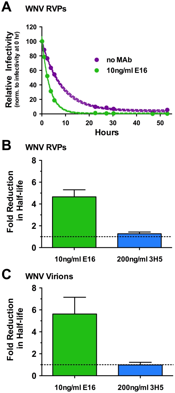
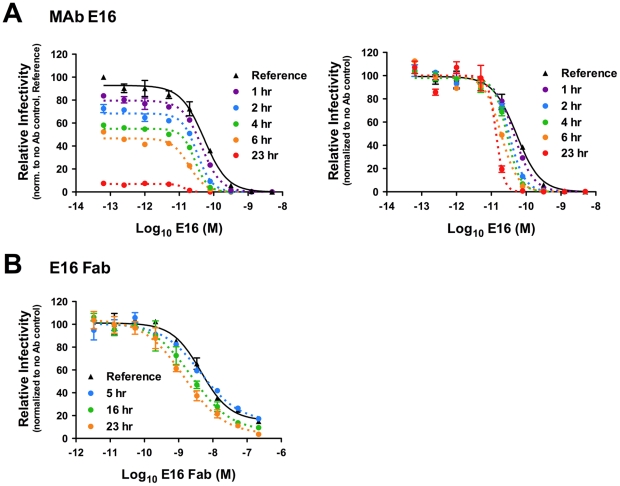
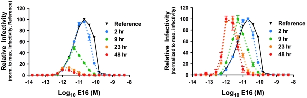
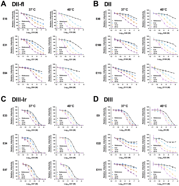
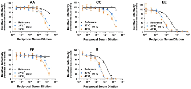
Neutralizing antibodies are a significant component of the host's protective response against flavivirus infection. Neutralization of flaviviruses occurs when individual virions are engaged by antibodies with a stoichiometry that exceeds a required threshold. From this "multiple-hit" perspective, the neutralizing activity of antibodies is governed by the affinity with which it binds its epitope and the number of times this determinant is displayed on the surface of the virion. In this study, we investigated time-dependent changes in the fate of West Nile virus (WNV) decorated with antibody in solution. Experiments with the well-characterized neutralizing monoclonal antibody (MAb) E16 revealed a significant increase in neutralization activity over time that could not be explained by the kinetics of antibody binding, virion aggregation, or the action of complement. Additional kinetic experiments using the fusion-loop specific MAb E53, which has limited neutralizing activity because it recognizes a relatively inaccessible epitope on mature virions, identified a role of virus "breathing" in regulating neutralization activity. Remarkably, MAb E53 neutralized mature WNV in a time- and temperature-dependent manner. This phenomenon was confirmed in studies with a large panel of MAbs specific for epitopes in each domain of the WNV envelope protein, with sera from recipients of a live attenuated WNV vaccine, and in experiments with dengue virus. Given enough time, significant inhibition of infection was observed even for antibodies with very limited, or no neutralizing activity in standard neutralization assays. Together, our data suggests that the structural dynamics of flaviviruses impacts antibody-mediated neutralization via exposure of otherwise inaccessible epitopes, allowing for antibodies to dock on the virion with a stoichiometry sufficient for neutralization.


Conflict of interest statement

The authors have declared that no competing interests exist.

Figures

Figure 1
Figure 1. The functional half-life of WNV decreases in the presence of virus-specific antibody.
(A) WNV RVPs were incubated in the absence or presence of sub-neutralizing concentrations (the EC50) of the MAb E16 for one hour at room temperature to allow binding to reach equilibrium. RVP-antibody complexes were then incubated at 37°C for the indicated times. The infectivity at each point was determined following infection of Raji-DC-SIGNR cells and monitored by flow cytometry 48 h post-infection. The data is presented normalized to levels obtained prior to incubation at 37°C (but after steady-state binding was reached) and fitted to a single-phase exponential decay to obtain the half-life. For the representative experiment shown, dotted lines represent 95% confidence intervals and error bars represent the standard error of duplicate measurements. The fold-reduction in the half-life of WNV RVPs (B) or infectious WNV (C) in the presence of sub-neutralizing quantities of MAb E16 (n = 23 and n = 2, respectively) or high concentrations of the DENV-specific MAb 3H5 (n = 2), as compared to the absence of antibody, are shown. Error bars represent the standard error.
Figure 2
Figure 2. Kinetic changes in antibody-mediated neutralization of WNV infection by the DIII-specific MAb E16.
Nine serial four-fold dilutions of MAb E16 (A) or E16 Fab fragments (B) were incubated with WNV RVPs for one hour at room temperature to allow binding to reach equilibrium. RVP-antibody complexes were then incubated at 37°C for incremental lengths of time before infecting Raji-DC-SIGNR cells. Infectivity was monitored by flow cytometry at 48 h post-infection. The reference curve represents RVP-antibody complexes added to target cells immediately after the room temperature incubation. For A, dose-response curves from a representative experiment are expressed relative to the infectivity of RVPs in the absence of antibody that were added to cells immediately after the room temperature incubation (left panel), or relative to the infectivity of RVPs in the absence of antibody at each individual time point (right panel). For B, the E16 Fab fragment results from a representative experiment are displayed relative to the infectivity of RVPs in the absence of antibody at each individual time point. Error bars display the standard error of duplicate infections. Results are representative of 19 and two independent experiments for A and B, respectively.
Figure 3
Figure 3. Kinetic changes in antibody dependent enhancement (ADE) of WNV infection by the MAb E16.
Nine serial four-fold dilutions of MAb E16 were incubated with WNV RVPs for one hour at room temperature to allow binding to reach equilibrium. RVP-antibody complexes were then incubated at 37°C for incremental lengths of time before infecting K562 cells. Infectivity was monitored by flow cytometry at 48 h post-infection. The reference curve represents RVP-antibody complexes added to target cells immediately after the room temperature incubation. Dose-response curves from a representative experiment are expressed relative to the maximum infectivity of RVPs that were added to cells immediately after the room temperature incubation (left panel), or relative to the maximum infectivity of RVPs at each individual time point (right panel). Error bars display the standard error of duplicate infections. Results are representative of four independent experiments.
Figure 4
Figure 4. Time- and temperature-dependent increases in neutralization by antibodies that bind cryptic determinants on the mature virion.
Nine serial four-fold dilutions of the DII-fusion loop (fl)-reactive MAb E53 (A and B) or the DI-reactive MAb E121 (C) were incubated with mature WNV RVPs for one hour at room temperature to allow binding to reach equilibrium. RVP-antibody complexes were then incubated at 37°C only (A) or at 33°C, 37°C, and 40°C (B and C) for incremental lengths of time before infecting Raji-DC-SIGNR cells. Infectivity was carried out at 37°C and monitored by flow cytometry at 48 h post-infection. The reference curve represents RVP-antibody complexes added to Raji-DC-SIGNR cells immediately after the room temperature incubation. Dose-response curves from representative experiments are expressed relative to the infectivity of RVPs in the absence of antibody at each individual time point. Error bars display the standard error of duplicate infections. Results are representative of five and three independent experiments for MAb E53 and E121, respectively.
Figure 5
Figure 5. Kinetic changes in neutralization occur for WNV MAbs specific for structurally distinct epitopes.
Nine serial four-fold dilutions of various WNV MAbs were incubated with mature WNV RVPs for one hour at room temperature to allow binding to reach equilibrium. RVP-antibody complexes were then incubated at 37°C or 40°C for incremental lengths of time before infecting Raji-DC-SIGNR cells. Infectivity was monitored by flow cytometry at 48 h post-infection. The reference curve represents RVP-antibody complexes added to Raji-DC-SIGNR cells immediately after the room temperature incubation. Dose-response curves are expressed relative to the infectivity of RVPs in the absence of antibody at each time point. Error bars display the standard error of duplicate infections. Results are representative of two independent experiments. MAbs selected for study were specific for epitopes on the DII-fusion loop (DII-fl) (A), the DII-central interface, DII-dimer interface, and DII-hinge interface (E48, E100, and E113, respectively) (B), the DIII-lateral ridge (DIII-lr) (C), and epitopes within DIII that fall outside of the lateral ridge (D).
Figure 6
Figure 6. Kinetic effects of WNV neutralization by polyclonal sera.
Nine serial three-fold dilutions of five heat-inactivated polyclonal WNV immune sera samples were incubated with mature WNV RVPs for one hour at room temperature to allow binding to reach equilibrium. RVP-antibody complexes were then incubated at 37°C or 40°C for incremental lengths of time before infecting Raji-DC-SIGNR cells. Infectivity was carried out at 37°C and monitored by flow cytometry at 48 h post-infection. The reference curve represents RVP-antibody complexes added to Raji-DC-SIGNR cells immediately after the room temperature incubation. Dose-response curves from a representative experiment are expressed relative to the infectivity in the absence of antibody at each individual time point. Error bars display the standard error of duplicate infections. Data is representative of two independent experiments.
Figure 7
Figure 7. A kinetic aspect of neutralization is also observed with DENV.
Nine serial four-fold dilutions of various DENV-1-reactive MAbs were incubated with DENV-1 RVPs for one hour at 37°C to allow binding to reach equilibrium. RVP-antibody complexes were then incubated at 37°C or 40°C for incremental lengths of time before infecting Raji-DC-SIGNR cells. Infectivity was carried out at 37°C and monitored by flow cytometry at 48 h post-infection. The reference curve represents RVP-antibody complexes added to Raji-DC-SIGNR cells immediately after the one hour incubation at 37°C. Dose-response curves from a representative experiment are expressed relative to the infectivity in the absence of antibody at each individual time point. Error bars display the standard error of duplicate infections. Data is representative of three (E95, E105) or two (E60, E102, E113) independent experiments. E95, E102, E105, and E113 are DIII-reactive MAbs specific for the G-strand, BC-loop, BC/DE/FG loops, and A-strand, respectively. E60 is a WNV-specific MAb that is cross-reactive for the DENV DII-fusion loop.

References

    1. Lindenbach BD, Thiel HJ, Rice CM. Flaviviridae: The Viruses and Their Replication. In: Knipe DM, Howley PM, editors. Fields Virology. 5th ed. Philadelphia: Lippincott-Williams & Wilkins; 2007. pp. 1101–1152.
    1. Sejvar JJ. The long-term outcomes of human West Nile virus infection. Clin Infect Dis. 2007;44:1617–1624. - PubMed
    1. Planitzer CB, Modrof J, Yu MY, Kreil TR. West Nile virus infection in plasma of blood and plasma donors, United States. Emerg Infect Dis. 2009;15:1668–1670. - PMC - PubMed
    1. Fernandez-Garcia MD, Mazzon M, Jacobs M, Amara A. Pathogenesis of flavivirus infections: using and abusing the host cell. Cell Host Microbe. 2009;5:318–328. - PubMed
    1. Brinton MA. The molecular biology of West Nile Virus: a new invader of the western hemisphere. Annu Rev Microbiol. 2002;56:371–402. - PubMed

Publication types