. 2011 Jun;7(6):e1002116.
doi: 10.1371/journal.ppat.1002116. Epub 2011 Jun 30.

The SV40 late protein VP4 is a viroporin that forms pores to disrupt membranes for viral release

Affiliations

The SV40 late protein VP4 is a viroporin that forms pores to disrupt membranes for viral release

Smita Raghava et al. PLoS Pathog. 2011 Jun.

Abstract

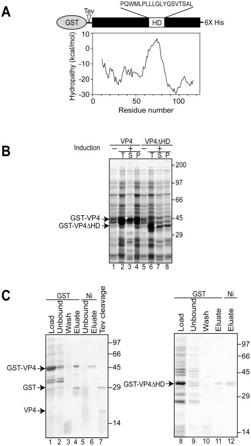
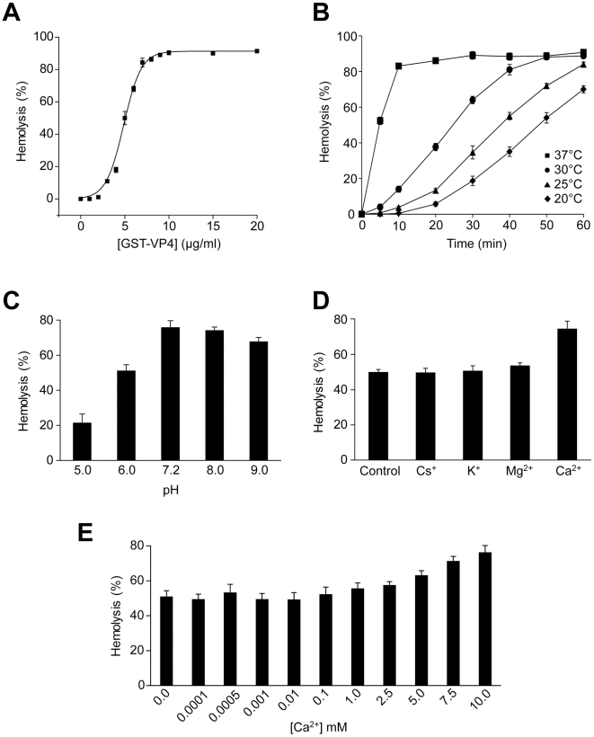
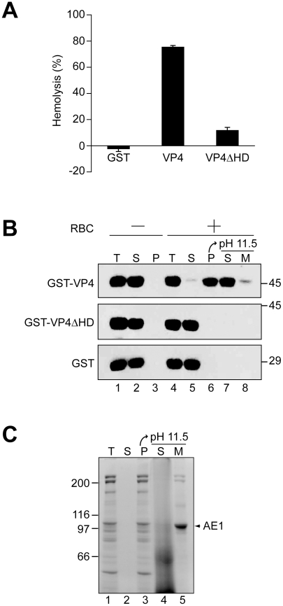
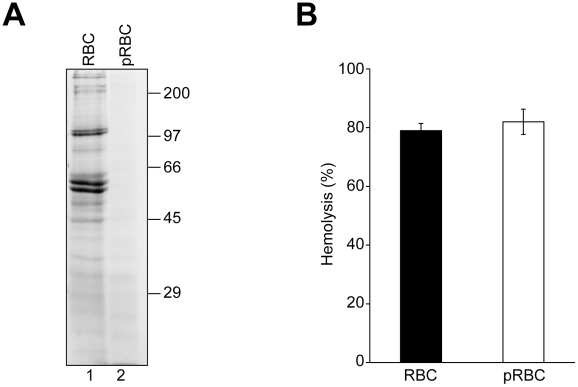
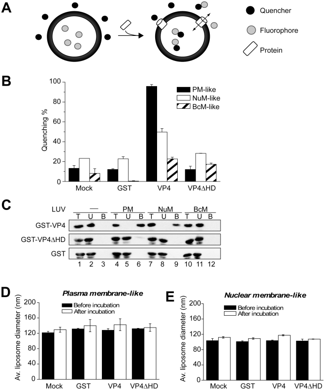
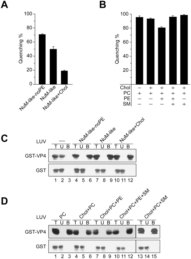
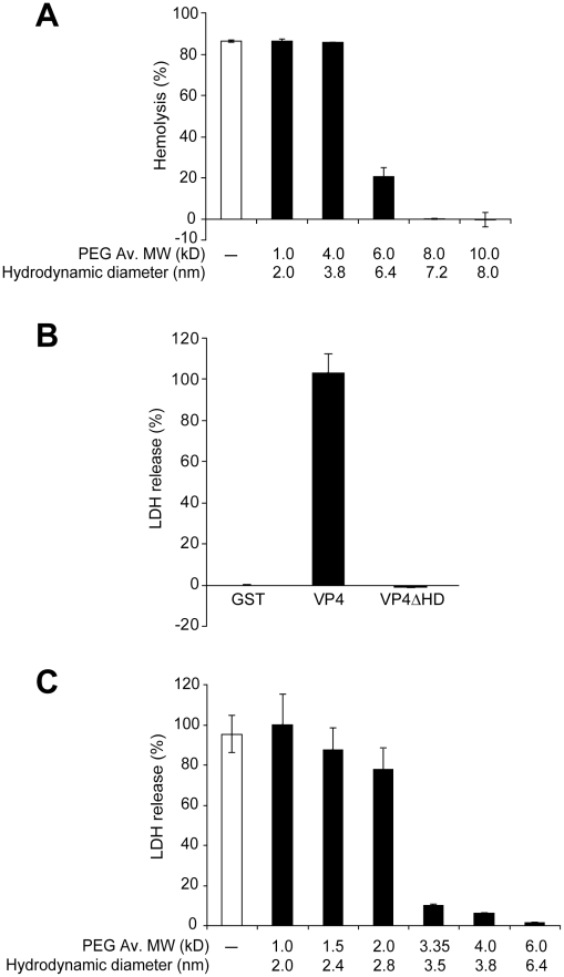
Nonenveloped viruses are generally released by the timely lysis of the host cell by a poorly understood process. For the nonenveloped virus SV40, virions assemble in the nucleus and then must be released from the host cell without being encapsulated by cellular membranes. This process appears to involve the well-controlled insertion of viral proteins into host cellular membranes rendering them permeable to large molecules. VP4 is a newly identified SV40 gene product that is expressed at late times during the viral life cycle that corresponds to the time of cell lysis. To investigate the role of this late expressed protein in viral release, water-soluble VP4 was expressed and purified as a GST fusion protein from bacteria. Purified VP4 was found to efficiently bind biological membranes and support their disruption. VP4 perforated membranes by directly interacting with the membrane bilayer as demonstrated by flotation assays and the release of fluorescent markers encapsulated into large unilamellar vesicles or liposomes. The central hydrophobic domain of VP4 was essential for membrane binding and disruption. VP4 displayed a preference for membranes comprised of lipids that replicated the composition of the plasma membranes over that of nuclear membranes. Phosphatidylethanolamine, a lipid found at high levels in bacterial membranes, was inhibitory against the membrane perforation activity of VP4. The disruption of membranes by VP4 involved the formation of pores of ∼3 nm inner diameter in mammalian cells including permissive SV40 host cells. Altogether, these results support a central role of VP4 acting as a viroporin in the perforation of cellular membranes to trigger SV40 viral release.


Conflict of interest statement

The authors have declared that no competing interests exist.

Figures

Figure 1
Figure 1. Bacterial expression and purification of VP4.
(A) Schematic representation of the VP4 construct containing N-terminal GST and C-terminal 6xHis tags. VP4 contains a hydrophobic domain (HD) with sequence designated. The hydrophobicity plot of VP4 using Membrane Protein Explorer version 3 is displayed. (B) Influence of the osmolyte, proline, on the solubility of GST-VP4 and GST-VP4ΔHD expressed in bacteria before (−) and after (+) induction with IPTG. Total (T), supernatant (S), pellet (P) fractions were resolved by SDS-PAGE and visualized by coomassie brilliant blue staining. Soluble GST-VP4 and GST-VP4ΔHD are designated by asterisks. (C) SDS-PAGE analysis of two-step affinity purification of GST-VP4 and GST-VP4ΔHD.
Figure 2
Figure 2. VP4 possesses hemolytic activity.
(A) Percent hemolysis of bovine RBCs as a function of GST-VP4 concentration. RBCs (0.5%, v/v) were incubated with various concentrations of the protein for 30 min at 37°C. (B) Kinetics of hemolysis of bovine RBCs as a function of temperature as indicated. RBCs were incubated at different temperatures with GST-VP4 (10 µg/ml) for 30 min. (C) Hemolysis as a function of pH. Hemolysis reactions were carried out at varying pH. Data are normalized with respect to samples containing only buffer and RBCs (0%), and containing RBCs with 50% water to mediate complete lysis (100%). (D) Hemolysis in presence of different metal ions. Purified GST-VP4 was mixed with bovine RBCs. Reactions were incubated in 10 mM Tris (pH 7.5)/150 mM NaCl containing Cs+, K+, Mg2+, Ca2+ ions. (E) Calcium concentrations were varied for the hemolytic assay over a wider range. The error bars represent the standard deviation from three independent experiments.
Figure 3
Figure 3. The hydrophobic domain of VP4 is required for its binding and disruption of RBC membranes.
(A) GST-VP4 or GST-VP4ΔHD were incubated with bovine RBCs for 30 min at 37°C. Released hemoglobin was measured by the A414 of the supernatant after centrifugation and the removal of unlysed cells. GST was used as a control to rule out its contribution in the hemolytic activity of GST-VP4. (B) Hemolysis reaction mixtures (lane 4, T) containing bovine RBCs and GST-VP4 or GST-VP4ΔHD were incubated at 37°C for 30 min. RBC bound (P, lane 6) and unbound (S, lane 5) proteins were separated by centrifugation. Membrane fractions (lane 6) were alkaline extracted with 0.1 M Na2CO3, pH 11.5 and ultracentrifuged to separate the soluble (S) and membrane (M) fractions (lane 7 and 8). Samples resolved by reducing SDS-PAGE were immunoblotted with antibody against GST. Separate reactions were performed in the absence of RBCs (lanes 1–3). (C) Bovine RBCs were separated as in B and the SDS-PAGE gel was stained with coomassie blue to visualize the abundant RBC membrane protein, anion exchanger 1 (AE1).
Figure 4
Figure 4. RBC surface proteins are not required for VP4-mediated hemolysis.
(A) RBCs (lane 1) were pretreated with trypsin and proteinase K (pRBCs, lane 2) to remove surface proteins. Membrane fractions after hypotonic lysis were resolved by reducing SDS-PAGE and proteins were visualized by coomassie blue staining. (B) GST-VP4 was mixed with untreated or protease treated RBCs. After incubation for 30 min at 37°C, hemoglobin release was measured by the A414 of the supernatant after centrifugation to remove unlysed cells. The error bars represent the standard deviation from two independent experiments performed in triplicate.
Figure 5
Figure 5. VP4 permeabilization of liposomes.
(A) Scheme showing the liposome disruption assay employed. Membrane disruption was examined by encapsulating [Tb(DPA)3- 3] fluorophore into LUVs (large unilamellar vesicles). When these LUVs were resuspended in a solution containing EDTA (quencher), protein mediated membrane disruption was monitored by the quenching of [Tb(DPA)3- 3] emission as the encapsulated molecules were released, and terbium ions were chelated by EDTA. (B) VP4 disrupts mammalian plasma (PM-like) and nuclear membrane-like (NuM-like) LUVs. LUVs mimicking the lipid compositions of bacterial inner membrane (BcM-like), and mammalian plasma and nuclear membranes were prepared to examine the membrane disruption activity of GST-VP4 and GST-VP4ΔHD. GST was used as a control. Mock LUVs were incubated in absence of any protein. Liposome disruption was evaluated using LUVs prepared with selected lipid compositions, and the percentage of fluorophore quenched is indicated. Each data point shows the average of at least two independent measurements with error bars representing standard deviations. (C) Flotation of proteins on sucrose gradients after incubation either without (-, lanes 1–3) or with PM-like (lanes 4–6), NuM-like (lanes 7–9), or BcM-like (lanes 10–12) LUVs to separate unbound (U; lanes 2, 5, 8 and 11) and bound (B; lanes 3, 6, 9 and 12) fractions. Proteins were resolved by SDS-PAGE and immunoblotting with anti-GST antibody. (D) Average diameter of PM-like LUVs before and after 30 min incubation with the indicated proteins as determined by dynamic light scattering. Each data point shows the average of at least two independent measurements with error bars representing standard deviation. (E) Average diameter of NuM-like LUVs determined similarly to D.
Figure 6
Figure 6. Activity of VP4 is dependent upon the lipid composition of the membranes.
(A) Membrane disruptive activity of GST-VP4 measured as percentage fluorescence quenching with two sets of LUVs where phosphatidylethanolamine (PE) was excluded from NuM-like LUVs (NuM-Iike-noPE) or the percentage of cholesterol was increased (NuM-Iike+Chol). Each data point shows the average of at least two independent measurements and the error bars denote the standard deviation of the experiment. See Table 1 for lipid fractions. (B) Membrane disruptive activity of GST-VP4 with five different sets of LUVs: PC, Chol+PC, Chol+PC+PE, Chol+PC+PE+SM and Chol+PC+SM. Each data point shows the average of at least two independent measurements and the error bars denote the standard deviation. (C) Flotation of proteins on sucrose gradients after incubation either without (-, lanes 1–3) or with NuM-like-noPE (lanes 4–6), NuM-like (lanes 7–9), or NuM-like+Chol (lanes 10–12) LUVs to separate unbound (U; lanes 2, 5, 8 and 11) and bound (B; lanes 3, 6, 9 and 12) fractions. Proteins were resolved by SDS-PAGE and immunoblotting with anti-GST antibody. (D) Flotation of proteins on sucrose gradients after incubation with PC (lanes 1–3), Chol+PC (lanes 4–6), Chol+PC+PE (lanes 7–9), Chol+PC+PE+SM (lanes 10–12), or Chol+PC+SM (lanes 13–15) LUVs. Proteins were resolved by SDS-PAGE and immunoblotting with anti-GST antibodies.
Figure 7
Figure 7. VP4 forms small size selective pores in biological membranes.
(A) Assessment of osmoprotection of red blood cells from lysis by VP4. Bovine RBCs (0.5%, v/v) were mixed with GST-VP4 (10 µg/ml) in the presence of polyethylene glycols (PEGs) of increasing molecular weights (1.0, 4.0, 6.0, 8.0 and 10.0 kD). The error bars represent the standard deviation from three independent experiments. (B) Percentage LDH released from Cos 7 cells after incubation with VP4 and VP4ΔHD for 30 min at 37°C. The percentage of LDH released was calculated by dividing the LDH released from samples by the LDH released with Triton X-100-permeabilized Cos 7 cells incubated under the same conditions (see Materials and Methods). The error bars represent the standard deviation from three independent experiments each carried out in triplicate. (C) Assessment of osmoprotection of Cos 7 cells from lysis by GST-VP4. Cos 7 cells were mixed with GST-VP4 (10 µg/ml) in the presence of different molecular weights PEGs (1.0, 1.5, 2.0, 3.35, 4.0 and 6.0 kD). The error bars represent the standard deviation from three independent experiments performed in triplicate.

References

    1. Chen BJ, Lamb RA. Mechanisms for enveloped virus budding: can some viruses do without an ESCRT? Virology. 2008;372:221–232. - PMC - PubMed
    1. Pornillos O, Garrus JE, Sundquist WI. Mechanisms of enveloped RNA virus budding. Trends Cell Biol. 2002;12:569–579. - PubMed
    1. Han Z, Harty RN. The NS3 protein of bluetongue virus exhibits viroporin-like properties. J Biol Chem. 2004;279:43092–43097. - PubMed
    1. Aldabe R, Barco A, Carrasco L. Membrane permeabilization by poliovirus proteins 2B and 2BC. J Biol Chem. 1996;271:23134–23137. - PubMed
    1. Fiers W, Contreras R, Haegemann G, Rogiers R, Van de Voorde A, et al. Complete nucleotide sequence of SV40 DNA. Nature. 1978;273:113–120. - PubMed

Publication types

MeSH terms