. 2011 Jun;7(6):e1002156.
doi: 10.1371/journal.pgen.1002156. Epub 2011 Jun 30.

Role of the single-stranded DNA-binding protein SsbB in pneumococcal transformation: maintenance of a reservoir for genetic plasticity

Affiliations

Role of the single-stranded DNA-binding protein SsbB in pneumococcal transformation: maintenance of a reservoir for genetic plasticity

Laetitia Attaiech et al. PLoS Genet. 2011 Jun.

Abstract

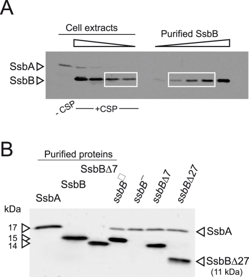
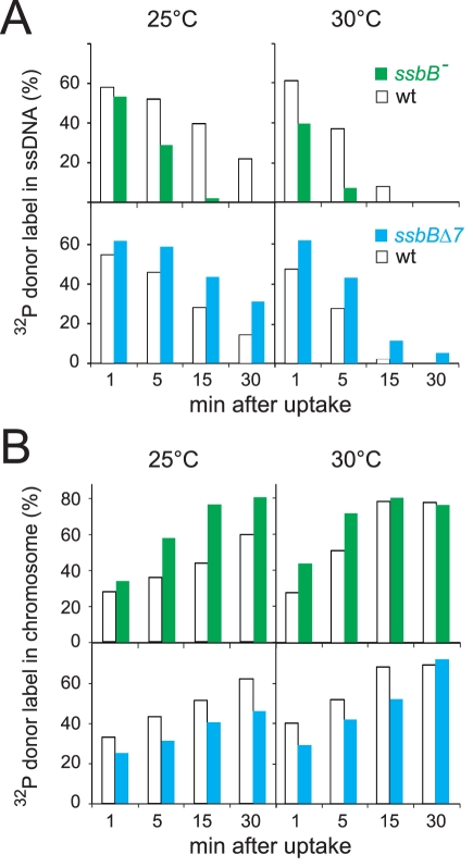
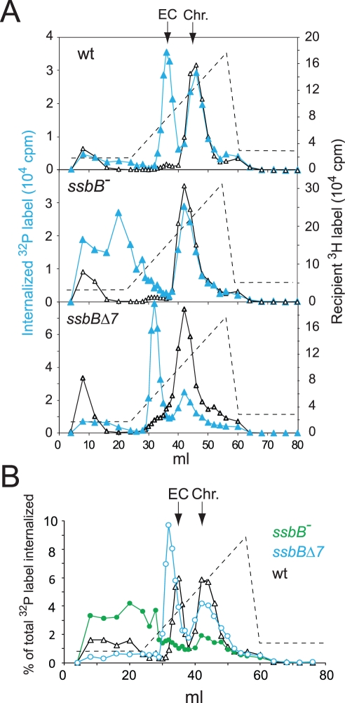
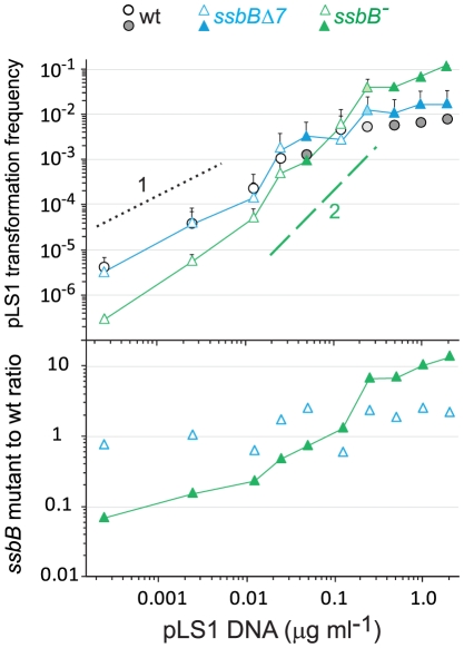
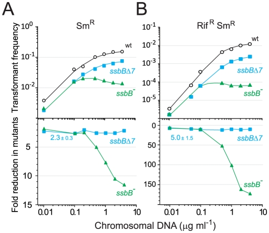
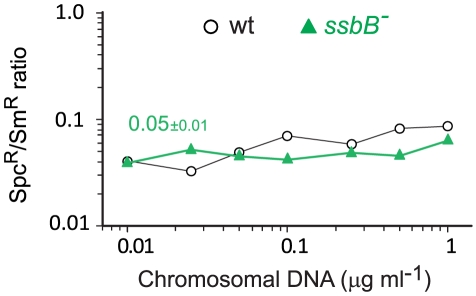
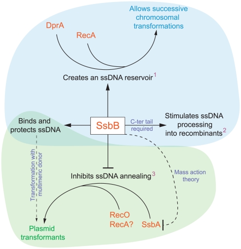
Bacteria encode a single-stranded DNA (ssDNA) binding protein (SSB) crucial for genome maintenance. In Bacillus subtilis and Streptococcus pneumoniae, an alternative SSB, SsbB, is expressed uniquely during competence for genetic transformation, but its precise role has been disappointingly obscure. Here, we report our investigations involving comparison of a null mutant (ssbB(-)) and a C-ter truncation (ssbBΔ7) of SsbB of S. pneumoniae, the latter constructed because SSBs' acidic tail has emerged as a key site for interactions with partner proteins. We provide evidence that SsbB directly protects internalized ssDNA. We show that SsbB is highly abundant, potentially allowing the binding of ~1.15 Mb ssDNA (half a genome equivalent); that it participates in the processing of ssDNA into recombinants; and that, at high DNA concentration, it is of crucial importance for chromosomal transformation whilst antagonizing plasmid transformation. While the latter observation explains a long-standing observation that plasmid transformation is very inefficient in S. pneumoniae (compared to chromosomal transformation), the former supports our previous suggestion that SsbB creates a reservoir of ssDNA, allowing successive recombination cycles. SsbBΔ7 fulfils the reservoir function, suggesting that SsbB C-ter is not necessary for processing protein(s) to access stored ssDNA. We propose that the evolutionary raison d'être of SsbB and its abundance is maintenance of this reservoir, which contributes to the genetic plasticity of S. pneumoniae by increasing the likelihood of multiple transformation events in the same cell.


Conflict of interest statement

The authors have declared that no competing interests exist.

Figures

Figure 1
Figure 1. Western blot analysis of S. pneumoniae SsbA and SsbB proteins.
(A) Quantification of SsbB in competent cells. For preparation of cell extracts, 20 ml culture in C+Y medium at OD550 0.13 (∼2.1 108 cfu ml−1) were divided into two equal parts, one of which received CSP (25 ng ml−1), and both were incubated for 12 min at 37°C. Total cells were then collected by centrifugation. Western blotting was as described in Materials and Methods. Volumes of total cell extracts, from left to right: 20 µl (−CSP) and 10, 5, 2.5 and 1.25 µl (+CSP). Amounts of purified SsbB, from left to right: 3.1, 6.3, 12.5, 25 and 50 ng. White rectangles identify cell extract samples and the corresponding purified protein standards used for SsbB quantification. SsbB amounts calculated for 2.5 and 1.25 µl extracts were 30.0 and 14.9 ng, respectively, resulting in an estimate of 70,799±300 SsbB molecules per cell, considering a cell density in the culture of ∼2.7 108 cells ml−1 (because ∼30% of daugther cells remain attached to each other after completion of cell division; our unpublished observations). (B) Western-blot analysis of competent ssbB +, ssbB , ssbBΔ7 and ssbBΔ27 cells. 5.3 µl of total extracts (corresponding to 235 µl culture at OD550 0.15) were analyzed as described above. Strains used: R1501 (ssbB +), R2294 (ssbB ), R2081 (ssbBΔ7) and R2082 (ssbBΔ27). Purified S. pneumoniae SsbA (50 ng), SsbB (200 ng) and SsbBΔ7 (200 ng) proteins were used as standard.
Figure 2
Figure 2. Fate of internalized ssDNA at 25°C and 30°C in wildtype and ssbB mutant cells.
(A) Kinetics of decay of internalized ssDNA. To investigate the impact of ssbB mutations on the stability of internalized ssDNA, pairwise comparison experiments involving an ssbB mutant (ssbB , R2201; or ssbBΔ7, R2204) and its wildtype parent (R1521) were conducted. Competent wildtype and ssbB mutant cells were transformed for 3 min with a 7771-bp S. pneumoniae fragment uniformly labeled with 32P, then incubated for 1, 5, 15 or 30 min; total DNA was extracted and the fate of transforming DNA was analyzed through agarose gel electrophoresis (Materials and Methods; Figure S2). The amount of ssDNA was calculated using densitometer tracings of electrophoregrams corresponding to extracts of cells transformed at 25°C (Figure S2, panels A and C; for an example, see blue rectangle in panel A), or in parallel at 30°C, and expressed relative to total donor DNA uptake in each extract; total uptake value for each time point was normalized to the average uptake value at 25°C (Figure S2B and S2D) or 30°C. (B) Kinetics of incorporation/integration of 32P donor label in the chromosome. Similarly to the measurement of ssDNA label described above, the amount of donor label incorporated/integrated into the chromosome was calculated from densitometer tracings of electrophoregrams, through integration of counts in the area corresponding to chromosomal DNA (for an example, see red rectangle in Figure S2A). Same symbols as in panel A.
Figure 3
Figure 3. Analysis of EC in wildtype, ssbB, and ssbBΔ7 cells.
(A) HAP chromatography of extracts from transformed wildtype (R2512), ssbBΔ7 (R2583) and ssbB (R2582) cells were run in parallel. Competent cells, pre-labeled with 3H-thymidine, were exposed for 5 min at 30°C to a mixture of unlabeled chromosomal DNA and a 32P-labeled 10,380-bp homologous fragment, then treated with DNase I for 1 min before lysis; preparation of cell extracts, chromatography on HAP and elution with a PB gradient were as described in Material and Methods. Blue symbols and lines, 32P donor label; black symbols and lines, 3H recipient label. Vertical arrows indicate the position of wildtype EC and chromosomal DNA (Chr.). Dotted line, PB gradient. (B) Duplicate (independent from panel A) HAP chromatography analyses of extracts from transformed wildtype (R2512), ssbBΔ7 (R2583) and ssbB (R2582) cells run in parallel. Recipient 3H chromosome label profiles have been omitted for clarity.
Figure 4
Figure 4. Differential effect of ssbB and ssbBΔ7 mutations on chromosomal and plasmid chromosomal transformation.
(A) Competent cells (∼1.5 108 cfu ml−1) of strain R1818 (ssbB +), R2647 (ssbBΔ7) and R2646 (ssbB ) were exposed to subsaturating concentrations (0.05 to 1 ng ml−1) of an 888-bp rpsL41 fragment. SmR transformants were scored. (B) Transformation with subsaturating concentrations of pLS1 plasmid DNA (0.00025 to 0.025 µg ml−1) using the same competent cells as in panel A. TcR transformants were scored.
Figure 5
Figure 5. SsbB antagonizes plasmid transformation at high DNA concentration.
Top panel: competent cells (∼1.1 108 cfu ml−1) of strain R1818 (ssbB +), R2647 (ssbBΔ7) and R2646 (ssbB ) were exposed to pLS1 plasmid DNA. TcR transformants were scored. Data are from five independent experiments with two different pLS1 DNA preparations (I and II, distinguished by open and color-filled symbols, respectively); transformation frequencies with 0.25 µg ml−1 DNA correspond to average of data obtained with the two preparations (light-coloured symbols). For clarity, only half error bars are figured. Dotted and dashed lines indicate, respectively, slope of 1 and 2 (i.e. linear and quadratic dependence on DNA concentration). Bottom panel: ratio of plasmid transformants between ssbB mutant and wildtype cells calculated from data in top panel.
Figure 6
Figure 6. Single and double transformation frequency in wildtype, ssbB, and ssbBΔ7 cells.
(A) Competent cells (∼3.0 108 cfu ml−1) of strain R1818 (ssbB +), R2647 (ssbBΔ7) and R2646 (ssbB ) were exposed to various concentrations of R304 chromosomal DNA and SmR transformants were scored. Data represent the compilation of six independent experiments shown in Figure S3 (left panel). Bottom panel: ratio between the number of SmR transformants in ssbB mutant and wildtype cells calculated from data in top panel. (B) Same conditions as in panel A but scoring for RifR SmR double transformants. Data represent the compilation of four independent experiments shown in Figure S3 (rigth panel). Bottom panel: ratio between the number of RifR SmR transformants in ssbB mutant and wildtype cells calculated from data in top panel.
Figure 7
Figure 7. Transformation of a 8,653–nt heterology and a point mutation in wildtype and ssbB cells.
Ratio between the number of SpcR (long heterology) and SmR (point mutation) transformants in strains R3055 (ssbB ) mutant and R1818 (wild type), calculated from data in Figure S4A and S4B).
Figure 8
Figure 8. Diagrammatic representation of the roles of SsbB in competent cells of S. pneumoniae.
SsbB binds and protects internalized ssDNA, which creates a reservoir (1at high donor DNA concentration) that can be accessed by DprA and RecA . This reservoir then allows successive chromosomal transformations in the same cell, which directly impacts on the generation of genetic diversity. SsbB also plays a direct, yet undetermined role in the processing of ssDNA into recombinants (2independently of the amount and length of ssDNA internalized), for which the C-ter tail of the protein is required. Finally, SsbB antagonizes plasmid transformation (3at high concentration of donor DNA). We suggest that while SsbA may interact with RecO and favor reconstitution of an intact plasmid replicon through RecO (or RecA) dependent single-strand annealing, SsbB does not because its Cter differs from that of SsbA; as SsbB is ∼20-fold more abundant than SsbA, mass action is likely to result in SsbB preferential binding, thus preventing annealing and therefore plasmid reconstitution. Chromosomal and plasmid transformation are distinguished respectively by blue and green zones.

References

    1. Hiller NL, Ahmed A, Powell E, Martin DP, Eutsey R, et al. Generation of genic diversity among Streptococcus pneumoniae strains via horizontal gene transfer during a chronic polyclonal pediatric infection. PLoS Pathog. 2010;6:e1001108. doi: 10.1371/journal.ppat.1001108. - DOI - PMC - PubMed
    1. Croucher NJ, Harris SR, Fraser C, Quail MA, Burton J, et al. Rapid pneumococcal evolution in response to clinical interventions. Science. 2011;331:430–434. - PMC - PubMed
    1. Lacks S, Greenberg B, Carlson K. Fate of donor DNA in pneumococcal transformation. J Mol Biol. 1967;29:327–347.
    1. Dagkessamanskaia A, Moscoso M, Hénard V, Guiral S, Overweg K, et al. Interconnection of competence, stress and CiaR regulons in Streptococcus pneumoniae: competence triggers stationary phase autolysis of ciaR mutant cells. Mol Microbiol. 2004;51:1071–1086. - PubMed
    1. Peterson S, Sung CK, Cline R, Desai BV, Snesrud E, et al. Identification of competence pheromone responsive genes in Streptococcus pneumoniae. Mol Microbiol. 2004;51:1051–1070. - PubMed

Publication types

MeSH terms

LinkOut - more resources