. 2011;6(6):e21341.
doi: 10.1371/journal.pone.0021341. Epub 2011 Jun 27.

Cloning and characterization of porcine 4Ig-B7-H3: a potent inhibitor of porcine T-cell activation

Affiliations

Cloning and characterization of porcine 4Ig-B7-H3: a potent inhibitor of porcine T-cell activation

Weiwei Chen et al. PLoS One. 2011.

Abstract

Background: Members of the B7 superfamily costimulate the proliferation of lymphocytes during the initiation and maintenance of antigen-specific humoral and cell-mediated immune responses. B7-H3 (CD276) is a newly identified member of the B7 superfamily. It has been shown that B7-H3 plays a significant role in regulating T cell response in humans and mice, but it is not known whether a counterpart of human or murine B7-H3 exists in porcine species.

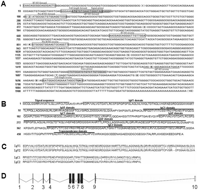
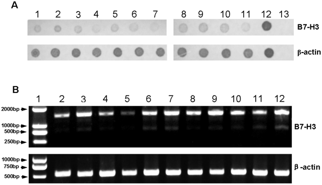
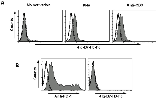
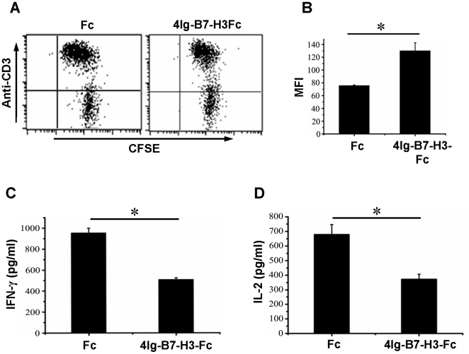
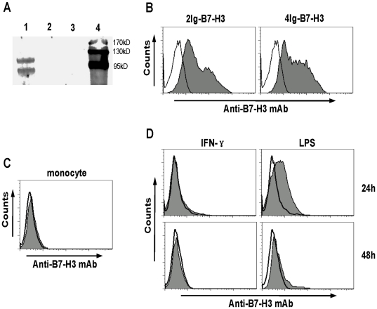
Methodology/principal findings: We cloned the porcine 4ig-b7-h3 gene using a blast search at the NCBI database with human b7-h3, RT-PCR and 3'-terminus RACE. Protein sequence analysis showed that the protein encoded by this gene contained 4Ig-like domains and was 90.88% identical with human 4Ig-B7-H3. Results of Dot-blot hybridization and RT-PCR showed that B7-H3 was broadly distributed in porcine tissues mainly as two isoforms, 2Ig-B7-H3 and 4Ig-B7-H3, of which 4Ig-B7-H3 was dominant. We further demonstrated that porcine 4Ig-B7-H3 was able to inhibit the proliferation and cytokine production of porcine T cells activated through the TCR pathway, similar to human B7-H3.

Conclusion: We cloned the porcine 4ig-b7-h3 gene and demonstrated that the porcine 4Ig-B7-H3 serves as a negative regulator for the T-cell immune response.


Conflict of interest statement

Competing Interests: This study was partly funded by HOUBIO TECH CO. LTD. There are no patents, products in development or marketed products to declare. This does not alter the authors' adherence to all the PLoS ONE policies on sharing data and materials.

Figures

Figure 1
Figure 1. Analysis of cDNA and the protein sequence of porcine 4Ig-B7-H3.
A, The cloned cDNA. The boxed, underlined and gray colored sequences show the positions of all used primers. The numbers in the brackets are the exon order of porcine B7-H3 in the genome. The strong ATG and TGA are initiation and stop codons. All exons contain the complete sequence in the genome but exon 1 in the cDNA. The numbers on the left are the base count of cDNA. B, The predicted protein sequence. The numbers on the left are the count of amino acids . All predicted domains are marked above the sequence. C, Alignment of the duplicated amino acid sequences of the two IgV and IgC-like domains, respectively. Asterisks indicate the identical amino acids. D, Schematic structure of CD276 nucleotide sequence in the porcine genome. The filled panes and lines represent Exons and Introns, respectively. The numbers under the schematics is the order of the Exons in the genome.
Figure 2
Figure 2. Distribution and analysis of the porcine B7-H3.
A, Dot-blot hybridization assay of mRNA from different tissues using the probes of 4Ig-B7-H3. Samples of PBMCs, PAM, heart, liver, spleen, lung, kidney, brain, lymph nodes, small intestine, tonsil were loaded from dot 1 to 11, respectively, with positive control (dot 12) and negative control (dot 13). The upper and lower panels were B7-H3 and β-actin, respectively. B, Analysis of the porcine B7-H3 isoforms. The normalized PCR products of PBMCs, PAM, heart, liver, spleen, lung, kidney, brain, lymph nodes, small intestine, tonsil were loaded from lane 2 to 12 with DNA ladder marker (lane 1). The upper and lower panels were the PCR products of B7-H3 and β-actin, respectively.
Figure 3
Figure 3. Analysis of a putative receptor of porcine 4Ig-B7-H3.
A, Binding of porcine 4Ig-B7-H3-Fc to T cells. T cells treated in different conditions were stained with a combination of anti-CD3 mAb with Fc (solid line histogram) or isotypic Ab (dotted line histogram) or 4Ig-B7-H3-Fc (filled histogram). Left plot: T cells in PBMC were not activated. Middle plot: T cells in primary PBMCs were stimulated with 5 µg/ml of PHA for 24 h. Right plot: T cells in primary PBMCs were stimulated with 1 µg/ml of anti-porcine CD3 mAb coated on the 96-well plate for 48 h. B, PD-1 gene was transfected into 293 T cells. PD-1 expressed on the 293 T cells was stained with anti-PD-1 mAb (left, filled histogram) or 4Ig-B7-H3-Fc protein (right, filled histogram) or Fc (solid line histogram) or isotypic Ab (dotted line histogram) 24 hours later.
Figure 4
Figure 4. Porcine 4Ig-B7-H3 inhibited T cell functions.
A, Freshly isolated porcine PBMCs were seeded to 96-well plate precoated with 1 µg/ml of anti-CD3 mAb plus either 10 µg/ml of 4Ig-B7-H3-Fc or Fc in 50 µl over night at 4°C. After culturing for 72 hours, T cell proliferation in the presence of Fc (left plot) or 4Ig-B7-H3-Fc (right plot) was analyzed. The plots were from one representative experiment of three independent samples. B, Statistic analysis of mean fluorescence intensity (MFI) on T cells (mean ± s.e.m). C and D, T cells purified from porcine PBMCs were seeded to the 96-well flat-bottomed plate precoated with 1 µg/ml of anti-CD3 mAb plus either 10 µg/ml 4Ig-B7-H3-Fc or Fc in 50 µl over night at 4°C. After culturing for 72 hours, supernatants were harvested to quantify IFN-γ (C) and IL-2 (D) concentration using ELISA kit. All data were from a representative of three independent experiments. * means P<0.05.
Figure 5
Figure 5. Analysis of porcine B7-H3 expression on monocytes.
A, Immunoprecipitation assay. The membrane protein extracted from the porcine 4Ig-B7-H3-transfected 293 T cells (Lane 1) or vacant plasmid-transfected 293 T cells (Lane 2) was pulled down with anti-porcine B7-H3 mAb-bound protein-G agarose. The pulled-down proteins were submitted to western-blot assay. Lane 3: anti-porcine B7-H3 mAb-bound protein-G agarose control. Lane 4: protein marker. B, The analysis of the porcine 2Ig-(left plot) and 4Ig-B7-H3 (right plot) expressed on 293 T cells stained by the anti-porcine B7-H3 mAb (filled histogram) or isotypic Ab (open histogram). C, non-activated monocytes were stained with a combination of anti-CD14 with either anti-B7-H3 mAb (filled histogram) or isotypic Ab (open histogram). D, The monocytes were co-cultured with IFN-γ in a final concentration of 30 ng/ml (left plots) or LPS at a final concentration of 1 µg/ml (right plots) for 24 h (upper plots) or 48 h plots (lower plots). On the indicated time points, the monocytes were harvested to be stained with a combination of anti-CD14 with either anti-B7-H3 mAb (filled histogram) or isotypic Ab (open histogram).

References

    1. Alegre ML, Frauwirth KA, Thompson CB. T-cell regulation by CD28 and CTLA-4. Nat Rev Immunol. 2001;1:220–228. - PubMed
    1. Dong C, Juedes AE, Temann UA, Shresta S, Allison JP, et al. ICOS co-stimulatory receptor is essential for T-cell activation and function. Nature. 2001;409:97–101. - PubMed
    1. Tafuri A, Shahinian A, Bladt F, Yoshinaga SK, Jordana M, et al. ICOS is essential for effective T-helper-cell responses. Nature. 2001;409:105–109. - PubMed
    1. Latchman Y, Wood CR, Chernova T, Chaudhary D, Borde M, et al. PD-L2 is a second ligand for PD-1 and inhibits T cell activation. Nat Immunol. 2001;2:261–268. - PubMed
    1. Selenko-Gebauer N, Majdic O, Szekeres A, Hofler G, Guthann E, et al. B7-H1 (programmed death-1 ligand) on dendritic cells is involved in the induction and maintenance of T cell anergy. J Immunol. 2003;170:3637–3644. - PubMed

Publication types