. 2011;6(6):e21699.
doi: 10.1371/journal.pone.0021699. Epub 2011 Jun 28.

A polymorphism in the splice donor site of ZNF419 results in the novel renal cell carcinoma-associated minor histocompatibility antigen ZAPHIR

Affiliations

A polymorphism in the splice donor site of ZNF419 results in the novel renal cell carcinoma-associated minor histocompatibility antigen ZAPHIR

Kelly Broen et al. PLoS One. 2011.

Abstract

Nonmyeloablative allogeneic stem cell transplantation (SCT) can induce remission in patients with renal cell carcinoma (RCC), but this graft-versus-tumor (GVT) effect is often accompanied by graft-versus-host disease (GVHD). Here, we evaluated minor histocompatibility antigen (MiHA)-specific T cell responses in two patients with metastatic RCC who were treated with reduced-intensity conditioning SCT followed by donor lymphocyte infusion (DLI). One patient had stable disease and emergence of SMCY.A2-specific CD8+ T cells was observed after DLI with the potential of targeting SMCY-expressing RCC tumor cells. The second patient experienced partial regression of lung metastases from whom we isolated a MiHA-specific CTL clone with the capability of targeting RCC cell lines. Whole genome association scanning revealed that this CTL recognizes a novel HLA-B7-restricted MiHA, designated ZAPHIR, resulting from a polymorphism in the splice donor site of the ZNF419 gene. Tetramer analysis showed that emergence of ZAPHIR-specific CD8+ T cells in peripheral blood occurred in the absence of GVHD. Furthermore, the expression of ZAPHIR in solid tumor cell lines indicates the involvement of ZAPHIR-specific CD8+ T cell responses in selective GVT immunity. These findings illustrate that the ZNF419-encoded MiHA ZAPHIR is an attractive target for specific immunotherapy after allogeneic SCT.


Conflict of interest statement

Competing Interests: The authors have declared that no competing interests exist.

Figures

Figure 1
Figure 1. Longitudinal follow-up of CD8+ T cells in peripheral blood from metastatic RCC patients UPN677 and UPN686 in relation to clinical outcome.
(A) UPN677: percentages of recipient T cells (right y axis) are compared with CD8+ T cell count ×106 per liter peripheral blood (left y axis). Administration of SCT and DLI 1–2 are indicated by ▴ and ▾, respectively. Treatment interval with CsA is shown by the dotted line. Time points of confirmed EBV infection, occurrence of EBV-specific T cells (depicted as % from total CD8+ T cell population) and death are indicated. (B) UPN686: for legend see (A). Time points of confirmed candidemia and death are indicated.
Figure 2
Figure 2. CD8+ T cells reactive with the SMCY.A2 peptide developed after DLI in RCC patient UPN677.
(A) HLA-restriction was determined by the production of IFN-γ released by CTL line H upon stimulation with recipient EBV-LCL in the presence of HLA blocking antibodies. Data are displayed as mean IFN-γ release ± SD of triplicate wells. (B) Production of IFN-γ by CTL line H stimulated with EBV-LCL of 9 HLA-A2+ unrelated individuals, showing recognition of 3 out of 9 EBV-LCL of male origin. (C) Flow cytometry analysis of CD8+ CTL line H simultaneously stained PE- and APC-conjugated SMCY.A2 tetramer, CD8 AlexaFluor 700, CD4, CD14, CD16 and CD19 FITC and Sytox Blue. CD8+ T cells were gated on FITC- and Sytox Blue- cells. The percentage of SMCY.A2 tetramer-binding cells among viable CD8+ T cells was 12.6%. These SMCY.A2 tetramer+ CD8+ T cells were sorted and expanded resulting in a >95% pure population. (D) Survival of the recipient and donor EBV-LCL, and donor EBV-LCL pulsed with peptide FIDSYICQV was determined in a flow cytometry-based cytotoxicity assay after incubation with SMCY.A2-specific CTL from patient UPN677 (▪), allo-HLA-A2 CTL (▾; positive control) or medium only (•) at an E:T ratio of 3∶1 in the presence of 25 U/ml IL-2. Data are depicted as mean ±SD of triplicate wells. (E) Detection of SMCY.A2-specific CD8+ T cells in peripheral blood of RCC patient UPN677. PBMC collected 24, 28 and 31 weeks post DLI-1 were simultaneously stained with PE- and APC-conjugated SMCY.A2 tetramer, CD8 AlexaFluor 700, CD4, CD14, CD16 and CD19 FITC and Sytox Blue. Subsequently, cell populations were analyzed by flow cytometry. Cells were gated on CD8+FITC-Sytox Blue- lymphocytes, and the percentage of tetramer-binding cells among CD8+ T cells is depicted.
Figure 3
Figure 3. Cytotoxicity of HLA-B7-restricted CTL B1 against RCC and hematological tumor cell lines.
(A) HLA-restriction was determined by the production of IFN-γ released by CTL B1 upon stimulation with recipient EBV-LCL in the presence of HLA blocking antibodies. (B) Production of IFN-γ by CTL B1 stimulated with rt EBV-LCL, EBV-LCL of an unrelated individual (#2) sharing HLA-B7 with the recipient, and an EBV-LCL of an HLA class I-mismatched individual (#3) that was transduced with HLA-B*0702. (C) Production of IFN-γ by CTL B1 and allo-HLA-B7 CTL stimulated with MiHA+ EBV-LCL and PHA-stimulated T cells and bone marrow-derived fibroblasts. Fibroblasts were pretreated with 10 ng/ml TNFα and 200 U/ml IFNγ for 2 days before coculture with CTL B1 (E:T ratio 10∶1). (D) MiHA mediated cytotoxicity on the HLA-B7+ RCC cell lines SKRC33 (rs2074071 genotype AA) and SKRC18 (rs2074071 genotype GG), the brain tumor cell line DAOY (rs2074071 genotype AG), and pharynx cell line FaDu (rs2074071 genotype GG), in flow cytometry-based cytotoxicity assays was determined after incubation with CTL B1 (▪), allo-HLA-B7 CTL (▾; positive control) or medium only (•) at an E:T ratio of 3∶1 in the presence of 25 U/ml IL-2. Data are depicted as mean ±SD of triplicate wells.
Figure 4
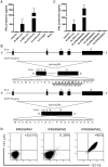
Figure 4. A SNP in the splice donor site between exon 4 and 5 of the ZNF419 gene determines CTL B1 recognition.
(A) IFN-γ production by CTL B1 upon stimulation with 293T-HLA-B*0702/ICAM/CD80 cells transfected with ZNF419 full length, cloned splice variant containing the exon4/exon5 boundary and the alternative ZNF419 splice variant containing intron 4. (B) Schematic representation of the ZNF419 gene on chromosome 19q13.43. The nucleotide and deduced amino acid sequences of the alternatively and correctly spliced transcripts present in recipient and donor, respectively, are shown. Disparity between the recipient and donor ZNF419 protein sequence is due to a G to A polymorphism in de splice donor site of intron 4 leading to the ZAPHIR epitope (underlined). (C) ZAPHIR epitope reconstitution with synthetic peptides corresponding to HLA-B7 10-mer peptide IPRDSWWVEL, 9-mer peptide IPRDSWWVE, 8-mer peptide IPRDSWWV, HLA-B7 10-mer peptide IPRGSWWVEL and HLA-B7 9-mer peptide IPRGEWHGA. Donor EBV-LCL was pulsed with 10 µM peptide and tested for recognition by CTL B1 in the presence of 25 U/ml IL-2. Data are displayed as mean ± SD of triplicate wells. (D) Flow cytometry analysis of CTL B1 with tetramers containing the 8-, 9-, or 10-mer peptide. Cells were stained with PE- and ACP-conjugated tetramer, CD8 AlexaFluor 700, CD4 FITC and Sytox Blue. Cells were analyzed by flow cytometry by gating on CD8+CD4-Sytox Blue- lymphocytes. The percentage of tetramer+ cells among CD8+ T cells is depicted.
Figure 5
Figure 5. ZNF419 microarray gene expression analysis of (malignant) hematopoietic and non-hematopoietic cells.
(A) ZNF419 expression of various hematopoietic cell types isolated from PBMC. (B) ZNF419 expression of hematopoietic malignancies sorted for CD34, CD33, and CD19/CD34. (C) ZNF419 expression of normal tissues +/− IFNγ. Data are displayed as MFIx106. 0–10 =  no expression, 10–100 =  low expression, 100–1000 =  intermediate expression, >1000 =  high expression.
Figure 6
Figure 6. Detection of ZAPHIR-specific CD8+ T cells in peripheral blood of RCC patient UPN686 and CML patient UPN539.
(A) PBMC collected before DLI and 4, 9, and 20 weeks post DLI were stained with PE- and ACP-conjugated ZAPHIR/HLA-B7 tetramer, CD8 AlexaFluor 700, CD4, CD14, CD16 and CD19 FITC and Sytox Blue. Cells were analyzed by flow cytometry by gating on CD8+FITC-Sytox Blue- lymphocytes. The percentage of tetramer+ cells among CD8+ T cells is depicted. The remaining PBMCs were stimulated once with ZAPHIR peptide pulsed (10 µM) EBV-LCL of the donor and assayed on day 7 for ZAPHIR-tetramer+ CD8+ T cells. (B) PBMC collected pre-DLI, 7 and 40 weeks post-DLI were stained and gated as described under (A). The remaining PBMCs were stimulated once with ZAPHIR peptide-pulsed (10 µM) EBV-LCL of the donor and assayed on day 7 for ZAPHIR-tetramer+ CD8+ T cells.

References

    1. Yang JC, Childs R. Immunotherapy for renal cell cancer. J Clin Oncol. 2006;24:5576–5583. - PubMed
    1. Childs R, Chernoff A, Contentin N, Bahceci E, Schrump D, et al. Regression of metastatic renal-cell carcinoma after nonmyeloablative allogeneic peripheral-blood stem-cell transplantation. N Engl J Med. 2000;343:750–758. - PubMed
    1. Pedrazzoli P, Da Prada GA, Giorgiani G, Schiavo R, Zambelli A, et al. Allogeneic blood stem cell transplantation after a reduced-intensity, preparative regimen - A pilot study in patients with refractory malignancies. Cancer. 2002;94:2409–2415. - PubMed
    1. Rini BI, Zimmerman T, Stadler WM, Gajewski TF, Vogelzang NJ. Allogeneic stem-cell transplantation of renal cell cancer after nonmyeloablative chemotherapy: feasibility, engraftment, and clinical results. J Clin Oncol. 2002;20:2017–2024. - PubMed
    1. Tykodi SS, Warren EH, Thompson JA, Riddell SR, Childs RW, et al. Allogeneic hematopoietic cell transplantation for metastatic renal cell carcinoma after nonmyeloablative conditioning: toxicity, clinical response, and immunological response to minor histocompatibility antigens. Clin Cancer Res. 2004;10:7799–7811. - PubMed

Publication types

Substances