. 2011;6(6):e21738.
doi: 10.1371/journal.pone.0021738. Epub 2011 Jun 29.

VapC toxins from Mycobacterium tuberculosis are ribonucleases that differentially inhibit growth and are neutralized by cognate VapB antitoxins

Affiliations

VapC toxins from Mycobacterium tuberculosis are ribonucleases that differentially inhibit growth and are neutralized by cognate VapB antitoxins

Bintou Ahmadou Ahidjo et al. PLoS One. 2011.

Abstract

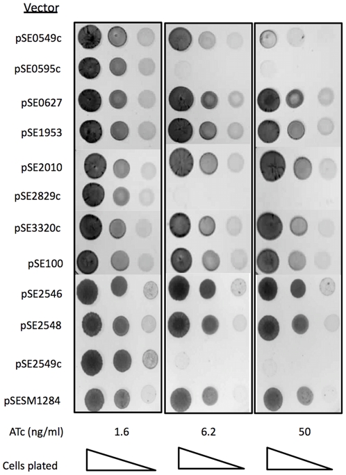
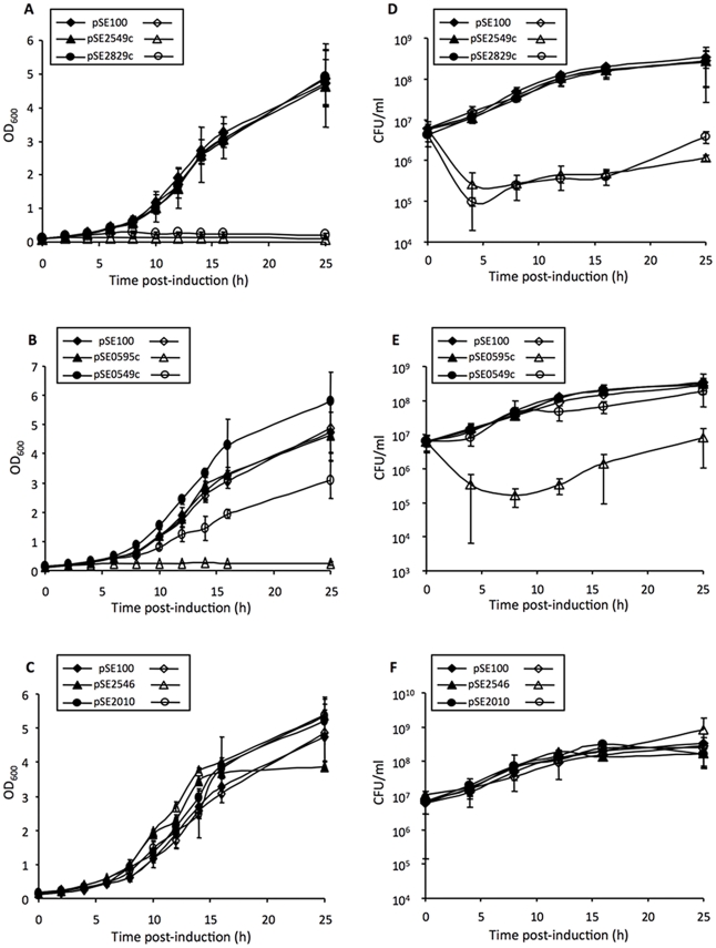
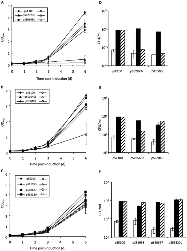
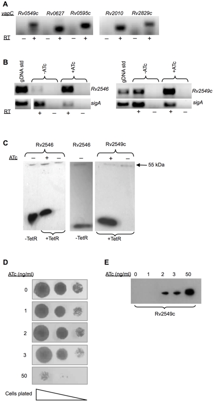
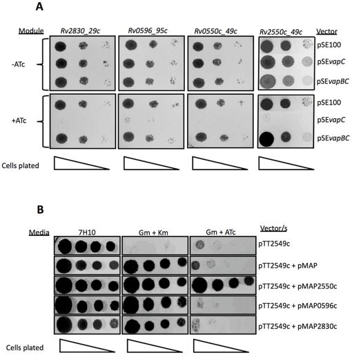
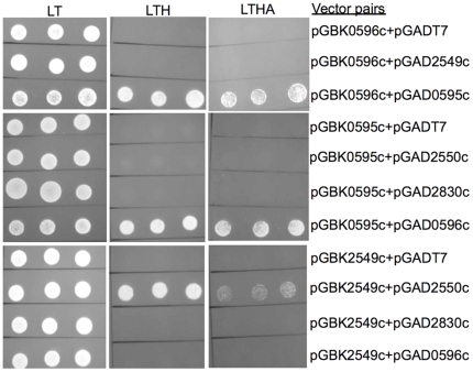
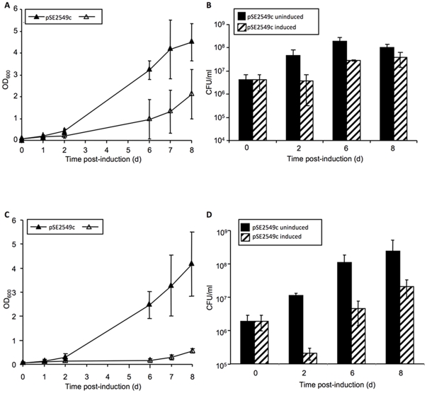
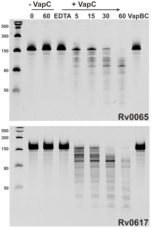
The chromosome of Mycobacterium tuberculosis (Mtb) encodes forty seven toxin-antitoxin modules belonging to the VapBC family. The role of these modules in the physiology of Mtb and the function(s) served by their expansion are unknown. We investigated ten vapBC modules from Mtb and the single vapBC from M. smegmatis. Of the Mtb vapCs assessed, only Rv0549c, Rv0595c, Rv2549c and Rv2829c were toxic when expressed from a tetracycline-regulated promoter in M. smegmatis. The same genes displayed toxicity when conditionally expressed in Mtb. Toxicity of Rv2549c in M. smegmatis correlated with the level of protein expressed, suggesting that the VapC level must exceed a threshold for toxicity to be observed. In addition, the level of Rv2456 protein induced in M. smegmatis was markedly lower than Rv2549c, which may account for the lack of toxicity of this and other VapCs scored as 'non-toxic'. The growth inhibitory effects of toxic VapCs were neutralized by expression of the cognate VapB as part of a vapBC operon or from a different chromosomal locus, while that of non-cognate antitoxins did not. These results demonstrated a specificity of interaction between VapCs and their cognate VapBs, a finding corroborated by yeast two-hybrid analyses. Deletion of selected vapC or vapBC genes did not affect mycobacterial growth in vitro, but rendered the organisms more susceptible to growth inhibition following toxic VapC expression. However, toxicity of 'non-toxic' VapCs was not unveiled in deletion mutant strains, even when the mutation eliminated the corresponding cognate VapB, presumably due to insufficient levels of VapC protein. Together with the ribonuclease (RNase) activity demonstrated for Rv0065 and Rv0617--VapC proteins with similarity to Rv0549c and Rv3320c, respectively--these results suggest that the VapBC family potentially provides an abundant source of RNase activity in Mtb, which may profoundly impact the physiology of the organism.


Conflict of interest statement

Competing Interests: The authors have declared that no competing interests exist.

Figures

Figure 1
Figure 1. Effect of mycobacterial VapC expression on growth of M. smegmatis on solid media.
Ten-fold serial dilutions of cells were spotted on 7H10 agar without or with ATc (1.6, 6.2 and 50 ng/ml) and incubated for 24–48 h.
Figure 2
Figure 2. Variable effects of Mtb VapC over-expression on growth and viability of M. smegmatis in liquid culture.
Growth and viability were assessed spectrophotometrically (A, B, C) and by CFU enumeration (D, E, F). Open symbols represent ATc-induced samples and filled symbols represent uninduced controls. The results represent the average and standard deviations from one of three independent experiments.
Figure 3
Figure 3. Variable effects of Mtb VapC expression on growth and viability of wild type Mtb in liquid culture.
A, B, C: Growth was assessed spectrophotometrically for a period of 6 days after induction of gene expression, as described under Materials and Methods. Open symbols represent ATc-induced samples and filled symbols represent uninduced controls. The results show the data from one of three independent experiments. D, E, F: Viability was assessed by enumerating CFU. Open bars, CFU prior to induction of gene expression (day 0); black bars, untreated control (day 3); and striped bars, ATc-treated culture (day 3). The results show the average CFU values from duplicate platings at one serial dilution from one of three independent experiments.
Figure 4
Figure 4. Analysis of ATc-regulated vapC expression in mycobacteria.
Expression was analyzed by RT-PCR (A and B) and detection of expressed proteins by epitope tagging (C). RT-PCR was carried out using mRNA samples obtained from (A) induced cultures of wild type Mtb (6 h treatment with ATc at 25 ng/ml); and (B) ATc-induced and uninduced cultures of M. smegmatis (1 h treatment with ATc at 50 ng/ml) over-expressing various vapC toxins under the control of the Pmyc1 tetO promoter. Samples obtained with (+) and without (-) reverse transcription (RT) were compared to distinguish cDNA from genomic DNA contamination. (C) Cellular fractions isolated from M. smegmatis were subjected to Western blot analysis using the anti-FLAG M2 antibody to detect the epitope-tagged VapC Rv2546 and Rv2549c proteins. Cultures were grown in either the presence (+) or absence (-) of 50 ng/ml ATc for 3 h. (D) The effect of epitope tagged Rv2549c expression on the growth of M. smegmatis on solid media. Ten-fold serial dilutions of cells were spotted on 7H10 agar without or with the ATc (1, 2, 3 and 50 ng/ml) and incubated for 24–48 h. (E) Comparison of the relative abundance of Rv2549c induced with varying concentrations of ATc. Cultures were grown in either the presence (+) or absence (−) of 50 ng/ml ATc for 3 h. Equal amounts (3 µg) were subjected to Western blot analysis using the anti-FLAG M2 antibody to detect the epitope-tagged Rv2549c protein.
Figure 5
Figure 5. Neutralization of Mtb VapC toxicity in M. smegmatis by cognate antitoxin expression.
A: Cognate toxin-antitoxin modules were co-expressed as an operon under control of the Tet-regulated promoter. Serial dilutions were plated on 7H10 agar alone (-ATc) or with ATc at 25 ng/ml (+ATc). B: Toxin and antitoxin genes were expressed separately on Tweety (vapC) or L5-based integration vectors (vapB) under control of Tet- or acetamide-regulated promoters, respectively. Serial dilutions were plated on 7H10 agar alone or supplemented with Gm and Km, or Gm and ATc (50 ng/ml).
Figure 6
Figure 6. Y2H analysis of VapC-VapB interactions.
Interactions between VapCs and either cognate or non-cognate VapBs were tested by co-transformation and scoring for growth on selection (LT) vs. media of differing stringency. Three independent colonies of each strain were re-suspended in sterile water, the cell density adjusted to an OD600 of 1 and aliquots spotted on plates. LT, Leu-Trp; LTH, Leu-Trp-His; LTHA, Leu-Trp-His-Ade dropout-supplemented media.
Figure 7
Figure 7. Rv2549c displays increased toxicity in the ΔRv2545-Rv2550c mutant strain of Mtb.
Growth and viability were assessed spectrophotometrically (A, C) and by CFU enumeration (B, D). Open symbols represent ATc-induced samples and filled symbols represent uninduced controls. The results represent the average and standard deviations from three independent experiments.
Figure 8
Figure 8. The VapCs Rv0065 and Rv0617 have RNase activity.
VapC proteins Rv0065 and Rv0617 display sequence-selective RNase activity against an RNA substrate of ∼150 bases in the presence of 6 mM MgCl2. Activity is Mg2+-dependent as addition of 12 mM EDTA abolishes activity (lane 4). Ribonuclease activity is also inhibited in the presence of VapB (lane 9). Addition of VapC (+VapC) results in degradation of the RNA substrate over a period of 5 – 60 minutes (lanes 5–8). RNA only controls (-VapC) show no contaminating ribonuclease activity (lanes 2 & 3). The molecular weight marker shows molecular masses of single-stranded RNA in number of bases (lane 1).

References

    1. Young DB, Perkins MD, Duncan K, Barry CE, 3rd Confronting the scientific obstacles to global control of tuberculosis. J Clin Invest. 2008;118:1255–1265. - PMC - PubMed
    1. Dhar N, McKinney JD. Microbial phenotypic heterogeneity and antibiotic tolerance. Curr Opin Microbiol. 2007;10:30–38. - PubMed
    1. Stewart GR, Robertson BD, Young DB. Tuberculosis: a problem with persistence. Nat Rev Microbiol. 2003;1:97–105. - PubMed
    1. Barry CE, 3rd, Boshoff HI, Dartois V, Dick T, Ehrt S, et al. Spectrum of latent tuberculosis - existing tests cannot resolve the underlying phenotypes: author's reply. Nat Rev Microbiol. 2010;8:242. - PubMed
    1. Warner DF, Mizrahi V. Tuberculosis chemotherapy: the influence of bacillary stress and damage response pathways on drug efficacy. Clin Microbiol Rev. 2006;19:558–570. - PMC - PubMed

Publication types

MeSH terms