. 2011 Mar-Apr;2(2):146-57.
doi: 10.4161/nucl.2.2.15211.

H3.3 is deposited at centromeres in S phase as a placeholder for newly assembled CENP-A in G₁ phase

Affiliations

H3.3 is deposited at centromeres in S phase as a placeholder for newly assembled CENP-A in G₁ phase

Elaine M Dunleavy et al. Nucleus. 2011 Mar-Apr.

Abstract

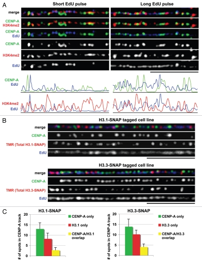
Centromeres are key regions of eukaryotic chromosomes that ensure proper chromosome segregation at cell division. In most eukaryotes, centromere identity is defined epigenetically by the presence of a centromeric histone H3 variant CenH3, called CENP-A in humans. How CENP-A is incorporated and reproducibly transmitted during the cell cycle is at the heart of this fundamental epigenetic mechanism. Centromeric DNA is replicated during S phase; however unlike replication-coupled assembly of canonical histones during S phase, newly synthesized CENP-A deposition at centromeres is restricted to a discrete time in late telophase/early G(1). These observations raise an important question: when 'old' CENP-A nucleosomes are segregated at the replication fork, are the resulting 'gaps' maintained until the next G(1), or are they filled by H3 nucleosomes during S phase and replaced by CENP-A in the following G(1)? Understanding such molecular mechanisms is important to reveal the composition/organization of centromeres in mitosis, when the kinetochore forms and functions. Here we investigate centromeric chromatin status during the cell cycle, using the SNAP-tag methodology to visualize old and new histones on extended chromatin fibers in human cells. Our results show that (1) both histone H3 variants H3.1 and H3.3 are deposited at centromeric domains in S phase and (2) there is reduced H3.3 (but not reduced H3.1) at centromeres in G(1) phase compared to S phase. These observations are consistent with a replacement model, where both H3.1 and H3.3 are deposited at centromeres in S phase and 'placeholder' H3.3 is replaced with CENP-A in G(1).

Keywords: CENP-A; DNA replication; cell cycle; centromere; histone deposition; kinetochore; mitosis.


Figures

Figure 1
Figure 1
Dilution and deposition of CENP-A visualized on chromatin fibers. (A) Chromatin fibers from S phase (EdU positive) or G1 phase (EdU negative, synchronized in mid-G1 by release from nocodazole block) cells stained with anti-CENP-A antibody. Cells were pulsed with EdU for 1 hour. DNA is stained with DAPI. Merge of CENP-A and DAPI is shown. Scale bar is 5 µM. (B) Quantitation of total CENP-A intensity measured using ImageJ (top) or total CENP-A spots (bottom) at centromeres in S phase (Cen S) compared to G1 phase (Cen G1), n=15 centromere fibers for each cell cycle stage (mean ± SD). (C) Newly synthesized CENP-A is deposited at centromeres in late telophase/early G1 phase. Cells expressing CENP-A-SNAP were labeled with TMR only (for total CENP-A-SNAP, top), or quenched with BTP and then immediately labeled with TMR with no chase time (to assess blocking efficiency, middle), or quenched with BTP then allowed 6 hours chase time for new protein synthesis before labeling of new CENP-A-SNAP with TMR (bottom). Cells were co-stained with anti-tubulin antibody that marks the midbody (white arrow) as an indicator of late telophase/early G1 phase cells. Scale bar is 15 µM. (D) (Upper part) Cell synchronization and experimental scheme for detection of newly synthesized CENP-A on chromatin fibers from G1 phase cells. (Lower part) Chromatin fibers from G1 cells were labeled with TMR to mark newly synthesized CENP-A-SNAP (red) and stained with anti-CENP-A antibody to mark total CENP-A (both total CENP-A-SNAP and endogenous CENP-A in green). 38 ± 13% of total CENP-A spots along the centromere track are new CENP-A-SNAP spots (n=20 fibers, mean ± SD). Scale bar is 5 µM.
Figure 2
Figure 2
Histone variants (H3.1 and H3.3) and histone modifications (H3K4me2) at centromeres in S phase. (A) Chromatin fibers showing replication of CENP-A and H3K4me2 domains at centromeres after a short (5 min) or long (5 h) pulse with EdU. Fibers are stained with anti-CENP-A antibody (green), anti-H3K4me2 antibody (red) and newly replicated DNA is marked with EdU (blue). Scale bar is 5 µM. Corresponding line plots illustrate the intensity of CENP-A (top) or H3K4me2 (bottom) with EdU along the centromere track after a short or long pulse with EdU. (B) Centromeric chromatin fibers from cells stably expressing H3.1-SNAP (top) and H3.3-SNAP (bottom). Total H3.1-SNAP or H3.3-SNAP is labeled with TMR (red), centromeres are marked with anti-CENP-A antibody (green), and newly replicated DNA is marked with EdU (blue, 1 hour pulse). Scale bar is 5 µM. (C) Quantitation shows that both H3.1-SNAP and H3.3-SNAP are mostly found in interspersed regions (red) between CENP-A and some overlap within CENP-A blocks (yellow), n=15 fibers for each cell line (mean ± SD).
Figure 3
Figure 3
New H3 deposition at centromeres in S phase. (A) Efficient ‘quenching’ of TMR (BTP, no chase, TMR) signal on whole cell nuclei in cells lines stably expressing H3.1-SNAP (left part) or H3.3-SNAP (right part). Anti-SNAP antibody (green) also recognizes SNAP that has been ‘quenched’ by BTP. Scale bar is 10 µM. (B) Experimental scheme to follow the incorporation of newly synthesized H3.1-SNAP or H3.3-SNAP on newly replicated DNA in S phase. (C) (Left part) On global non-centromeric chromatin, newly synthesized H3.1-SNAP is deposited on EdU positive fibers and is absent from EdU negative fibers. (Right part) On global chromatin, newly synthesized H3.3-SNAP is deposited on both EdU positive fibers and EdU negative fibers. Scale bar is 5 µM, n=20 fibers in each case. (D) Deposition of newly synthesized H3.1-SNAP at centromeres in S phase. Centromeres are marked with anti-CENP-A antibody (green), new H3.1-SNAP with TMR (red), newly replicated DNA with EdU (blue) and DNA is stained with DAPI. Merge of TMR, CENP-A and EdU is shown. Scale bar is 5 µM. Quantification (n = 16 fibers, mean ± SD) of the domain where new H3.1-SNAP is deposited reveals that new H3.1-SNAP is deposited at CENP-A domains (yellow, black arrows) or interspersed domains (red) with equal frequencies. (E) Deposition of newly synthesized H3.3-SNAP at centromeres in S phase. Centromeres are marked with anti-CENP-A antibody (green), new H3.3-SNAP with TMR (red), newly replicated DNA with EdU (blue) and DNA is stained with DAPI. Merge of TMR, CENP-A and EdU is shown. Scale bar is 5 µM. Quantification (n=16 fibers, mean ± SD) of the domain where new H3.3-SNAP is deposited reveals that new H3.3-SNAP is deposited more frequently in interspersed domains (red) than CENP-A domains (yellow, black arrows).
Figure 4
Figure 4
Total H3.3 at centromeres is reduced in G1 compared to S phase. (A) Experimental scheme for TMR labeling of total H3.1-SNAP or H3.3-SNAP at centromeres in S or G1 phase cells. (B) Chromatin fibers showing total H3.3-SNAP (TMR) at centromeres (upper part), or global chromatin (lower part) in S phase (EdU positive), or G1 phase (EdU negative). Centromeres are marked with anti-CENP-A antibody (green), total H3.3-SNAP is labeled with TMR (red), newly replicated DNA with EdU and DNA is stained with DAPI (gray). Scale bar is 5 µM. (C) Quantitation of total H3.3-SNAP (left part) or H3.1-SNAP (right part) intensity on chromatin fibers from centromeres or global chromatin in S and G1 cell cycle phases. Total TMR intensity on centromere fibers marked by CENP-A staining (cen S or cen G1) or on a non-centromeric fiber of equal length (global S or global G1) was measured using ImageJ, n=16 fibers for each data set. The amount of H3.3-SNAP at centromeres drops significantly in G1 (EdU negative) compared to S phase (EdU positive) (p=0.000038, Student's t test). (D) Quantitation of CENP-A spots (green), H3.3 spots (red) or CENP-A/H3.3 spots (yellow) at centromeres in S and G1 phases, n=16 fibers, mean ± SD. The number of H3.3 spots and CENP-A/H3.3 spots at centromeres is reduced in G1 phase compared to S phase.
Figure 5
Figure 5
Model for the dynamics of CENP-A, H3.1 and H3.3 deposition at centromeres during the cell cycle. CENP-A blocks at centromeres are interspersed with H3.1 blocks and H3.3 blocks (each circle is one nucleosome). During S phase, parental (old) CENP-A nucleosomes are redistributed to daughter strands and are diluted by half. Newly synthesized H3.1 and H3.3 are deposited as ‘placeholders’ at CENP-A domains and interspersed domains. Half of new H3.1 is deposited at CENP-A domains and half at interspersed domains, whereas new H3.3 is more frequently deposited at interspersed domains than at CENP-A domains. After S phase, half of H3.1 at centromeres is new, whereas most H3.3 at centromeres is newly deposited. During G2 phase or mitosis H3.3 may be removed. During G1 phase ‘placeholder’ H3.3 is replaced by new CENP-A. The amount of H3.3 at centromeres during G1 is reduced compared to S phase, whereas H3.1 does not change. New CENP-A deposition may not always occur at the same centromeric position during every cell cycle.

References

    1. Allshire RC, Karpen GH. Epigenetic regulation of centromeric chromatin: Old dogs, new tricks? Nat Rev Genet. 2008;9:923–937. - PMC - PubMed
    1. Shelby RD, Monier K, Sullivan KF. Chromatin assembly at kinetochores is uncoupled from DNA replication. J Cell Biol. 2000;151:1113–1118. - PMC - PubMed
    1. Jansen LE, Black BE, Foltz DR, Cleveland DW. Propagation of centromeric chromatin requires exit from mitosis. J Cell Biol. 2007;176:795–805. - PMC - PubMed
    1. Shelby RD, Vafa O, Sullivan KF. Assembly of CENP-A into centromeric chromatin requires a cooperative array of nucleosomal DNA contact sites. J Cell Biol. 1997;136:501–513. - PMC - PubMed
    1. Sullivan KF. A solid foundation: Functional specialization of centromeric chromatin. Curr Opin Genet Dev. 2001;11:182–188. - PubMed

Publication types