. 2011 Sep;83(9):1571-81.
doi: 10.1002/jmv.22133.

Molecular determinants of disease in coxsackievirus B1 murine infection

Affiliations

Molecular determinants of disease in coxsackievirus B1 murine infection

Javier O Cifuente et al. J Med Virol. 2011 Sep.

Abstract

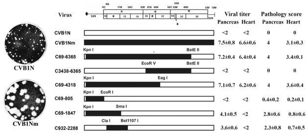
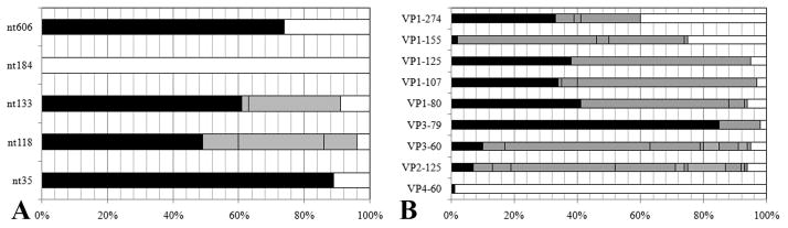
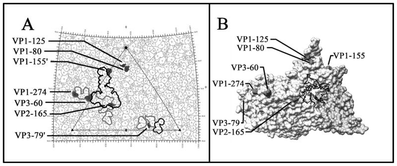
To understand better how different genomic regions may confer pathogenicity for the coxsackievirus B (CVB), two intratypic CVB1 variants, and a number of recombinant viruses were studied. Sequencing analysis showed 23 nucleotide changes between the parental non-pathogenic CVB1N and the pathogenic CVB1Nm. Mutations present in CVB1Nm were more conserved than those in CVB1N when compared to other CVB sequences. Inoculation in C3H/HeJ mice showed that the P1 region is critical for pathogenicity in murine pancreas and heart. The molecular determinants of disease for these organs partially overlap. Several P1 region amino acid differences appear to be located in the decay-accelerating factor (DAF) footprint CVBs. CVB1N and CVB1Nm interacted with human CAR, but only CVB1N seemed to interact with human DAF, as determined using soluble receptors in a plaque-reduction assay. However, the murine homolog Daf-1 did not interact with any virus assessed by hemagglutination. The results of this study suggest that an unknown receptor interaction with the virus play an important role in the pathogenicity of CVB1Nm. Further in vivo studies may clarify this issue.


Figures

Figure 1
Figure 1. Schematic representation of CVB1 parental and recombinant viruses, viral titres and pathology score
The restriction sites used to obtain the constructs are noted above the margins of the chimeric segments. CVB1N segments are shown as open bars while CVB1Nm segments are shown as solid bars. The pathology score was obtained as described in the text. Viral titres are expressed as log10 TCID50±SE per gram of tissue; <2 indicates that the infectivity was below the limit of the sensitivity assay. All determinations were performed using tissues corresponding to the C3H/HeJ mice shown in Figure 4.
Figure 2
Figure 2. Sequencing studies
A: Bar chart showing frequency of identical nucleotides (including gaps) of the 5′UTR (A) and identical amino acid residues for the capsid proteins (B) from alignments with other human enterovirus B (taxid:138949). The frequency of matching identity is shown for CVB1Nm (black), CVB1N (white), with unmatched identity indicated in grey.
Figure 3
Figure 3. Comparative studies on HeLa cells
One-step growth curves with a multiplicity of infection (MOI) of 10 for each virus were performed on HeLa cells. At the indicated time points after infection, cells were washed with 0.1 ml of PBS, harvested and freeze-thawed three times. The live viral particles were counted using a plaque-forming assay. P<0.05 after comparison of CVB1N and C932-2288 with CVB1Nm and the remaining recombinant viruses. p.i.= post-inoculation.
Figure 4
Figure 4. Representative histology of murine pancreas and heart after inoculation with CVB1N, CVB1Nm and intratypic recombinant viruses
Ten days after inoculation of weanling male C3H/HeJ with these viruses, the pancreas and heart were harvested and processed for routine staining with haematoxylin and eosin. Representative sections from the pancreas (right panels) and myocardium (left panels) of mice inoculated with CVB1N (A–B), CVB1Nm (C–D), C69-4318 (E–F), C69-805 (G–H), C69-1847 (IJ), and C932-2288 (K–L) are shown. Scale bars: 125 μm.
Figure 5
Figure 5
A: The viral surface is represented as a quilt of amino acids and shown as a stereographic projection, where the polar angles θ and ϕ represent latitude and longitude, respectively [Xiao and Rossmann, 2006]. The viral icosahedral asymmetric unit is indicated by the triangular boundary. The DAF footprint for echoviruses (a grey outline) was plotted using the equivalent CVB3 residues based on multiple sequence alignments [Chenna et al., 2003] according to the DAF contact residues for EV7 [He et al., 2002] and EV12 [Bhella et al., 2004]. EV and CVB3 have a sequence identity of 61% for VP1, 70% for VP2 and 67% for VP3. The DAF footprint on CVB3 is outlined in black. Mutations are indicated in black. VP1-155 and VP3-79 are highlighted in neighbouring asymmetric units. Five of the seven amino acid changes, which map to the viral surface, are located within areas of CVB3 that are known to interact with DAF. ‘ Denotes symmetry-related amino acids. B: This figure was obtained using Chimera [Pettersen et al., 2004; Sanner et al., 1996]. One protomer of the CVB3 surface rendered with a symmetry-related copy of VP3 to show the canyon and CAR binding region, relative to the location of the CVB1Nm surface residue changes. None of the mutations are within the CAR binding site. The closest residue, VP2-165, is located opposite the puff at a distance from the canyon.

References

    1. Altschul SF, Madden TL, Schaffer AA, Zhang J, Zhang Z, Miller W, Lipman DJ. Gapped BLAST and PSI-BLAST: a new generation of protein database search programs. Nucleic Acids Res. 1997;25:3389–3402. - PMC - PubMed
    1. Beck MA, Levander OA, Handy J. Selenium deficiency and viral infection. J Nutr. 2003;133:1463S–1467S. - PubMed
    1. Belnap DM, McDermott BM, Jr, Filman DJ, Cheng N, Trus BL, Zuccola HJ, Racaniello VR, Hogle JM, Steven AC. Three-dimensional structure of poliovirus receptor bound to poliovirus. Proc Natl Acad Sci USA. 2000;97:73–78. - PMC - PubMed
    1. Bergelson JM, Cunningham JA, Droguett G, Kurt-Jones EA, Krithivas A, Hong JS, Horwitz MS, Crowell RL, Finberg RW. Isolation of a common receptor for Coxsackie B viruses and adenoviruses 2 and 5. Science. 1997;275:1320–1323. - PubMed
    1. Bergelson JM, Mohanty JG, Crowell RL, St John NF, Lublin DM, Finberg RW. Coxsackievirus B3 adapted to growth in RD cells binds to decay-accelerating factor (CD55) J Virol. 1995;69:1903–1936. - PMC - PubMed

Publication types

Substances