. 2012 May;22(5):1188-201.
doi: 10.1002/hipo.20964. Epub 2011 Jul 7.

4- to 6-week-old adult-born hippocampal neurons influence novelty-evoked exploration and contextual fear conditioning

Affiliations

4- to 6-week-old adult-born hippocampal neurons influence novelty-evoked exploration and contextual fear conditioning

Christine A Denny et al. Hippocampus. 2012 May.

Abstract

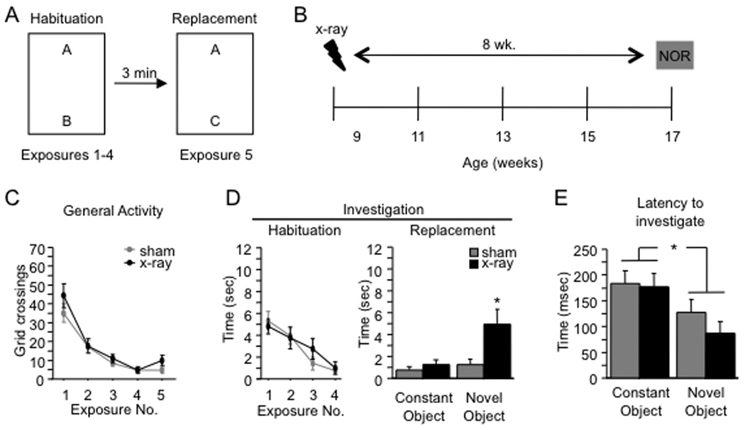
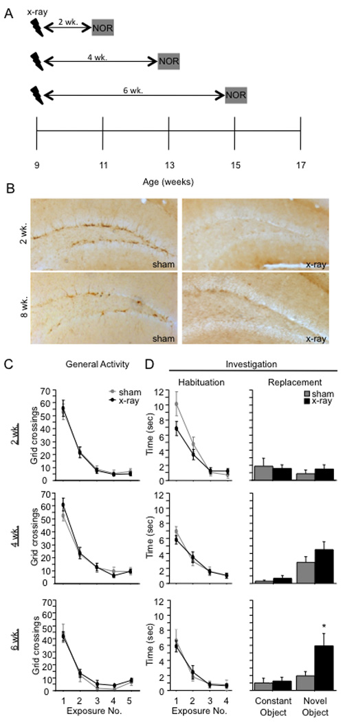
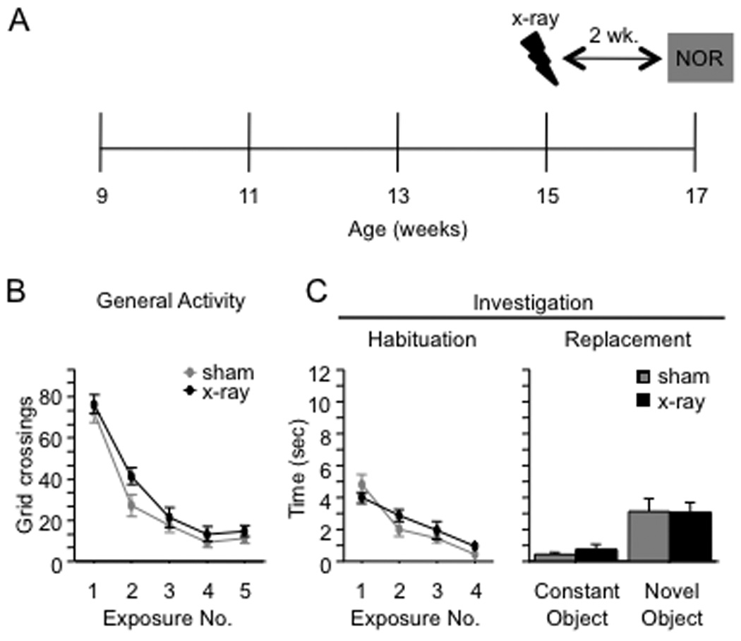
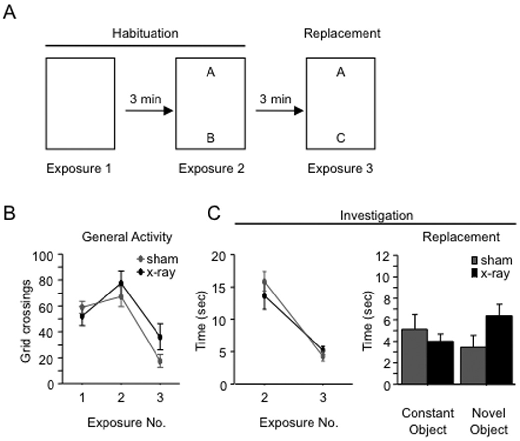
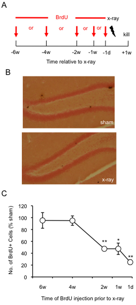
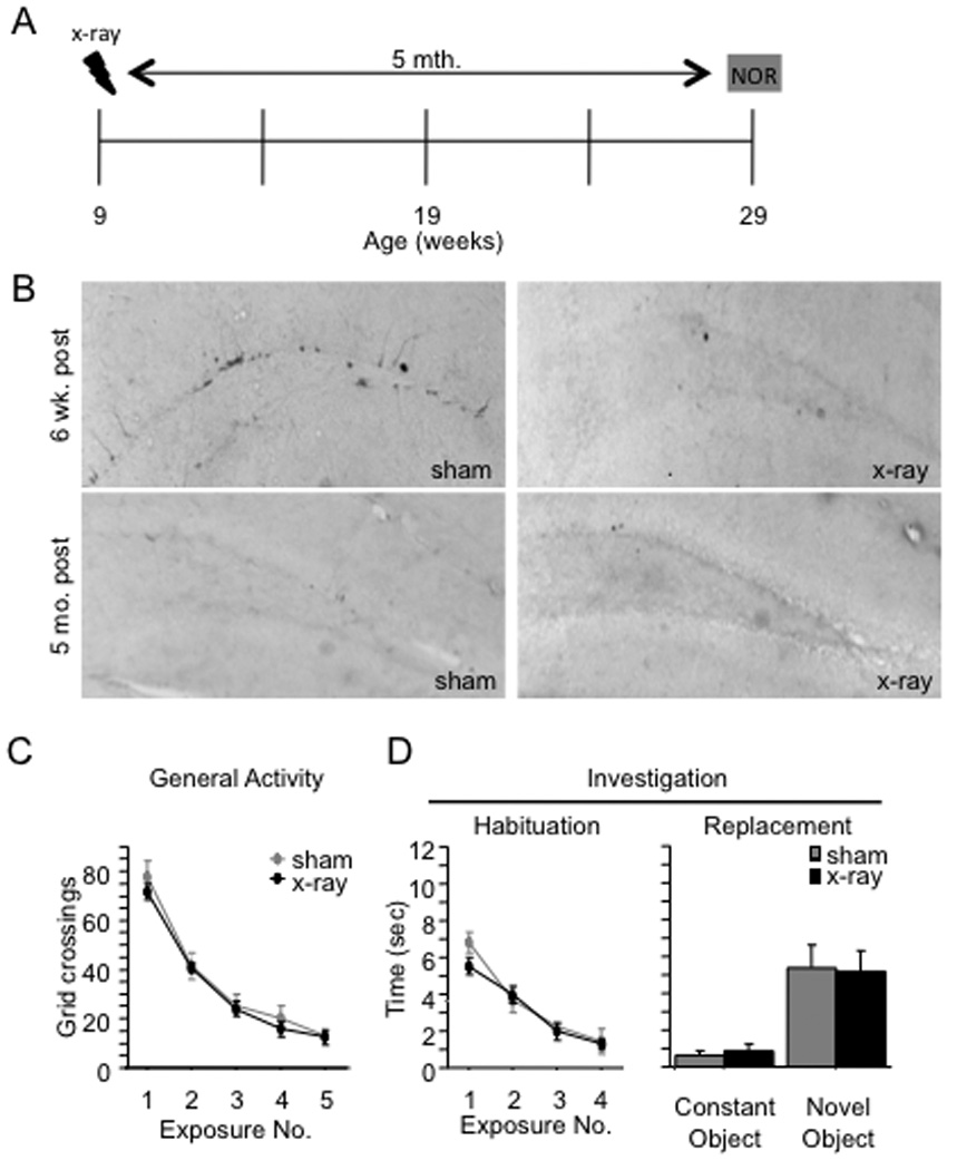
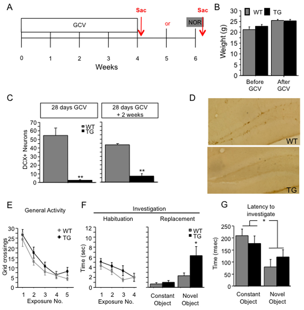
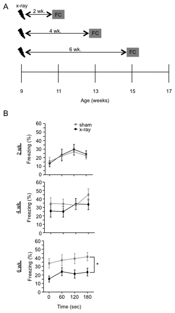
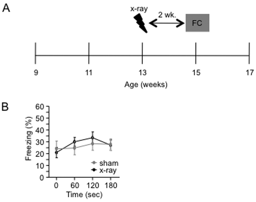
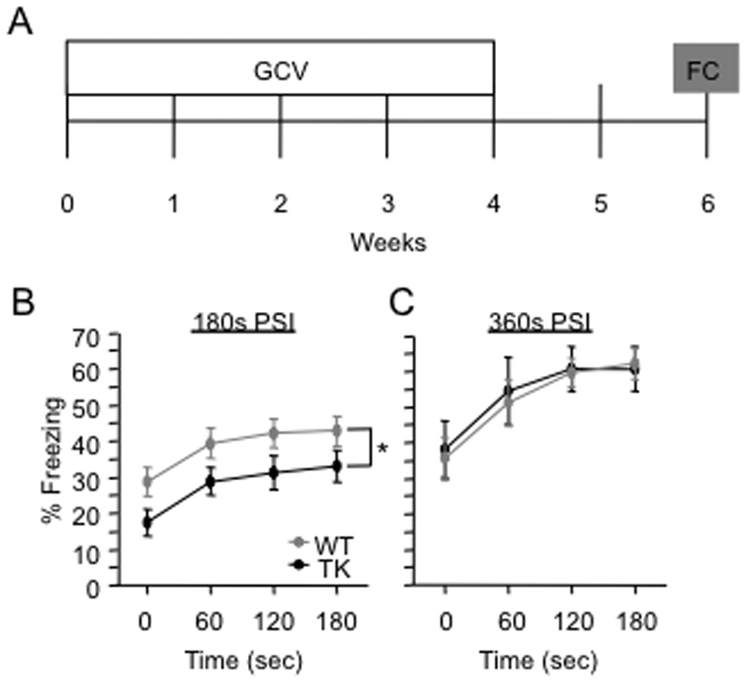
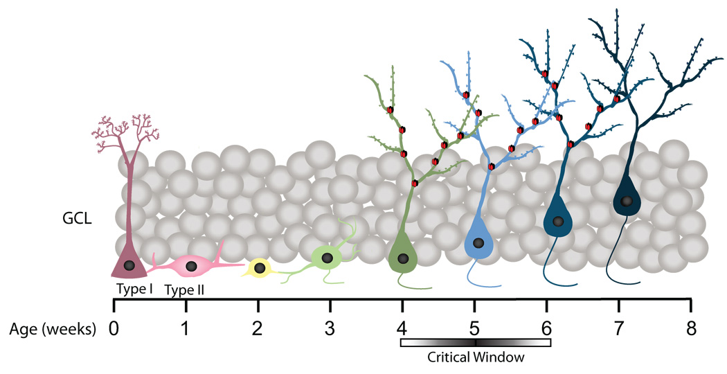
To explore the role of adult hippocampal neurogenesis in novelty processing, we assessed novel object recognition (NOR) in mice after neurogenesis was arrested using focal x-irradiation of the hippocampus, or a reversible, genetic method in which glial fibrillary acidic protein-positive neural progenitor cells are ablated with ganciclovir. Arresting neurogenesis did not alter general activity or object investigation during four exposures with two constant objects. However, when a novel object replaced a constant object, mice with neurogenesis arrested by either ablation method showed increased exploration of the novel object when compared with control mice. The increased novel object exploration did not manifest until 4-6 weeks after x-irradiation or 6 weeks following a genetic ablation, indicating that exploration of the novel object is increased specifically by the elimination of 4- to 6-week-old adult born neurons. The increased novel object exploration was also observed in older mice, which exhibited a marked reduction in neurogenesis relative to young mice. Mice with neurogenesis arrested by either ablation method were also impaired in one-trial contextual fear conditioning (CFC) at 6 weeks but not at 4 weeks following ablation, further supporting the idea that 4- to 6-week-old adult born neurons are necessary for specific forms of hippocampal-dependent learning, and suggesting that the NOR and CFC effects have a common underlying mechanism. These data suggest that the transient enhancement of plasticity observed in young adult-born neurons contributes to cognitive functions.


Figures

Figure 1
Figure 1. X-irradiation increases novel object investigation
(A) Schematic diagram of the NOR paradigm. Mice received 4 exposures to two objects. For exposure 5, one of the objects was replaced with a novel object. (B) Mice were x-irradiated or sham-irradiated at 9 weeks of age and the NOR paradigm was administered 8 weeks later at 17 weeks of age. (C–D) General activity and investigation (habituation; averaged across both objects) declined across exposures 1–4 for both x-irradiated and sham mice. There was no effect of x-irradiation on either variable [F’s(1,19) < 1]. In exposure 5 (replacement), x-irradiated mice investigated the novel object more than sham mice (p = 0.02), but the groups did not differ in exploration of the constant object (p = 0.42). General activity during exposure 5 was not affected by x-irradiation (p = 0.11). (F) The latency to investigate the novel object was shorter than the latency to investigate the constant object for both groups of mice (p < 0.01), and the latencies did not differ between groups (p = 0.36). * p < 0.05. ** p < 0.01. Error bars represent ± SEM.
Figure 2
Figure 2. The x-irradiation-induced increase in novel object investigation has a delayed onset
(A) Schematic diagram of the experimental time-course. (B) DCX immunoreactivity is absent 2 and 8 weeks following x-irradiation, demonstrating that neurogenesis was arrested at all points during behavioral testing. (C–D) At 2 weeks post x-irradiation, there was no effect of x-irradiation on any parameter. At 4 and 6 weeks post x-irradiation, x-irradiated and sham mice exhibited similar levels of general activity and object investigation in exposures 1–4 [F’s < 1]. At 4 weeks post x-irradiation, x-irradiated mice appeared to investigate the novel object more than sham mice during exposure 5, but the effect did not reach significance [F(1,20) = 2.95, p = 0.10]. At 6 weeks following x-irradiation, x-irradiated mice explored the novel object significantly more than sham mice during exposure 5 ([t(13) = 2.2, p = 0.047]). * p < 0.05. Error bars represent ± SEM.
Figure 3
Figure 3. NOR performance is related to the length of time after x-irradiation rather than the absolute age of the mouse
(A) Schematic diagram of the experimental time-course. (B–C) X-irradiated and sham mice did not differ in general activity or investigation (habituation) during exposures 1 through 4 [F’s(1,18) < 2.4, p’s > 0.14]. Both groups of mice investigated the novel object more than the constant object [F(1,22) = 24.3, p < 0.001], and there was no effect of the x-irradiation treatment [F’s(1,22) < 1]. Error bars represent ± SEM.
Figure 4
Figure 4. Pre-exposure to the NOR arena fails to increase novel object preference
(A) Schematic diagram of the experimental time-course. Mice were pre-exposed to the arena before introduction of the objects. (B–C) Pre-exposure to the arena increased overall levels of object investigation in both groups of mice when a novel object was introduced; however, both groups explored the constant and novel object similarly (n = 6–8 mice/group). Error bars represent ± SEM.
Figure 5
Figure 5. Hippocampal x-irradiation targets adult-born neurons 2-weeks old and younger
(A) Schematic diagram of the experimental BrdU injection protocol. (B) Representative images of the DG of sham and x-irradiated mice injected with BrdU at 2 weeks before x-irradiation and processed for BrdU immunoreactivity. (C) Mean number BrdU+ cells expressed as percent of sham mice. Mean number of BrdU+ cells is significantly lower in x-irradiated mice when compared with sham mice at 2 w, 1 w, and 1 d before x-irradiation. * p < 0.05, ** p < 0.01. Error bars represent ± SEM.
Figure 6
Figure 6. Aging is associated with a decline in hippocampal neurogenesis and an increase in novel object exploration
(A) Schematic diagram of the experimental time-course. (B) While DCX immunoreactivity is present in sham mice at 6 weeks post x-irradiation, DCX immunoreactivity is absent in sham mice at 5 months post x-irradiation. DCX immunoreactivity is abolished in x-irradiated mice at 6 weeks and 5 months post x-irradiation, indicating a permanent ablation of hippocampal neurogenesis. (C–D) In exposures 1 through 5, x-irradiated and sham mice exhibited similar levels of general activity [F(1,18) < 1, p = 0.596] and investigation [F(1,18) < 1, p = 0.39]. Both groups of mice investigated the novel object more than the constant object, and there was no effect of x-irradiation on investigation of either object. Error bars represent ± SEM.
Figure 7
Figure 7. GCV treatment increases novel object exploration in GFAP-TK TG mice
(A) Schematic diagram of the experimental time-course. Mice were treated with GCV for 28 days and then were tested in the NOR paradigm 2 weeks later. (B) GCV treatment did not induce body weight loss as indicated by comparable body weights between GFAP-TK TG and WT mice before and after GCV treatment. (C) DCX+ young neurons were significantly reduced in GFAP-TK TG mice treated with GCV when compared with WT mice, indicating that GCV had significantly reduced neurogenesis. (D) Representative images of the DG processed for DCX immunoreactivity. (E–F) During exposures 1 through 4, WT and GFAP-TK TG mice exhibited similar levels of general activity [F(1,44) = 1.4, p = 0.25] and investigation [F(1,44) = 1.3, p = 0.255]. In exposure 5, GFAP-TK TG mice explored the novel object more than WT mice [t(44) = 2.1, p = 0.041], but the groups did not differ in investigation of the constant object (p = 0.453). (G) The latency to the novel object was shorter than that to the constant object for both groups of mice (p < 0.01). There was no effect of genotype on the latency to either object (p = 0.87). * p < 0.05. ** p < 0.01. Error bars represent ± SEM.
Figure 8
Figure 8. The effect of x-irradiation on 1-trial contextual fear conditioning has a delayed onset
(A) Schematic diagram of the experimental time-course. Mice were fear conditioned 2, 4, or 6 weeks after x-irradiation. Contextual fear conditioning was produced by placing a mouse in the conditioning chamber and delivering one footshock 180 s later. Mice were returned to the conditioning chamber 24 h later to assess for context-elicited freezing. (B) Context-elicited freezing was significantly reduced in x-irradiated mice when compared with sham mice at 6 weeks following x-irradiation [F(1,32) = 7.1, p = 0.0119] but not at 2 or 4 weeks following x-irradiation [F’s(1,18) < 1]. * p < 0.05. Error bars represent ± SEM.
Figure 9
Figure 9. 1-trial contextual fear conditioning performance is related to the length of time after x-irradiation rather than the absolute age of the mouse
(A) Schematic diagram of the experimental time course. Mice were x-irradiated at 13 weeks of age and then fear conditioned two weeks later. (B) X-irradiated and sham mice did not differ in the test of context-elicited fear conducted 24 h following fear conditioning [F(1,27) < 1, p = 0.77]. Error bars represent ± SEM.
Figure 10
Figure 10. GCV treatment impairs 1-trial contextual fear conditioning in GFAP-TK TG mice
(A) Schematic diagram of the experimental time-course. GFAP-TK TG and WT mice were treated with GCV for 4 weeks and then fear conditioned 2 weeks later. Contextual fear conditioning was produced by placing a mouse in the conditioning chamber and delivering one footshock at 180 s or at 360 s later. In the test of context-elicited fear 24 h following training, GFAP-TK TG mice exhibited significantly less freezing than WT mice when the placement to shock interval (PSI) was 180 s (B) [F(1,66) = 4.0, p = 0.0498]. However, when the PSI was 360 s (C), GFAP-TK TG mice exhibited similar levels of freezing as WT mice in the test of context-elicited fear conducted 24 h following training [F(1,15) < 1, p = 0.8682]. * p < 0.05. Error bars represent ± SEM.
Figure 11
Figure 11. A schematic summary of neuronal differentiation in the dentate gyrus
Cells aged 4–6 weeks old modulate NOR and CFC performance. At this time point, adult-born neurons have unique features that enhance plasticity, such as elevated expression of NR2B-containing NMDA receptors (Ge et al., 2007). NR2B-containing NMDA receptors are denoted as red and black subunits.

References

    1. Aggleton JP, Keen S, Warburton EC, Bussey TJ. Extensive cytotoxic lesions involving both the rhinal cortices and area TE impair recognition but spare spatial alternation in the rat. Brain Res Bull. 1997;43:279–287. - PubMed
    1. Gray A. The neuropsychology of anxiety: an enquiry into the functions of the septo-hippocampal system. Oxford: Oxford University Press; 1982. p. 548.
    1. Baxter MG, Murray EA. Opposite relationship of hippocampal and rhinal cortex damage to delayed nonmatching-to-sample deficits in monkeys. Hippocampus. 2001:61–71. - PubMed
    1. Beason-Held LL, Rosene DL, Killiany RJ, Moss MB. Hippocampal formation lesions produce memory impairment in the rhesus monkey. Hippocampus. 1999;9:562–574. - PubMed
    1. Ben Abdallah NM-B, Slomianka L, Vyssotski AL, Lipp H-P. Early age-related changes in adult hippocampal neurogenesis in C57 mice. Neurobiol Aging. 2010;31:151–161. - PubMed

Publication types