. 2012 Jul;33(7):1607-20.
doi: 10.1002/hbm.21311. Epub 2011 Jul 7.

Cortical thickness is linked to executive functioning in adulthood and aging

Affiliations

Cortical thickness is linked to executive functioning in adulthood and aging

Agnieszka Z Burzynska et al. Hum Brain Mapp. 2012 Jul.

Abstract

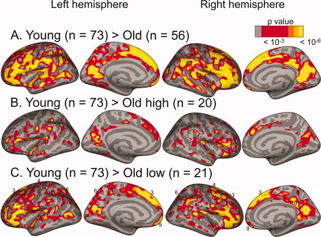
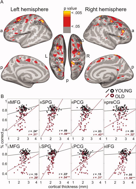
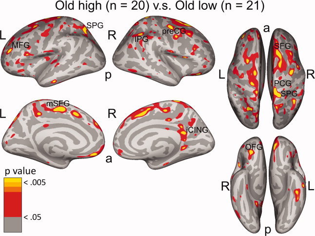
Executive functions that are dependent upon the frontal-parietal network decline considerably during the course of normal aging. To delineate neuroanatomical correlates of age-related executive impairment, we investigated the relation between cortical thickness and executive functioning in 73 younger (20-32 years) and 56 older (60-71 years) healthy adults. Executive functioning was assessed using the Wisconsin Card Sorting Test (WCST). Cortical thickness was measured at each location of the cortical mantle using surface-based segmentation procedures on high-resolution T1-weighted magnetic resonance images. For regions involved in WCST performance, such as the lateral prefrontal and parietal cortices, we found that thicker cortex was related to higher accuracy. Follow-up ROI-based analyses revealed that these associations were stronger in older than in younger adults. Moreover, among older adults, high and low performers differed in cortical thickness within regions generally linked to WCST performance. Our results indicate that the structural cortical correlates of executive functioning largely overlap with previously identified functional patterns. We conclude that structural preservation of relevant brain regions is associated with higher levels of executive performance in old age, and underscore the need to consider the heterogeneity of brain aging in relation to cognitive functioning.


Figures

Figure 1
Figure 1
Age‐group comparisons show a strong and spatially distributed negative effect of age on cortical thickness. Red to yellow: regions where cortex was thicker in younger than in (A) older adults, (B) high‐performing older adults, and (C) low‐performing older adults. In (C) numbers indicate, 1: left frontal pole (BA 10), 2: caudal part of the MFG (BA 6/8), 3: lateral superior frontal gyrus (BA 6/8/9), 4: precentral gyrus (BA 4), 5: rostral part of the MFG (BA9/46), 6: posterior parietal cortex (BA 7), 7: left caudal superior temporal gyrus (BA 22), 8: medial postcentral gyrus (BA 5), 9: orbitofrontal cortex (BA 11), and 10: right isthmus cingulate cortex (BA 23).
Figure 2
Figure 2
Relationship between cortical thickness and executive performance. A: Thicker cortical mantle was associated with better WCST performance (red‐yellow) in the total sample (corrected for age), and B: Mean cortical thickness from regions at P < 0.005 larger than 30 mm2 in panel A regressed on WCST accuracy. x‐axis: cortical thickness in mm, y‐axis: WCST accuracy (% correct responses); r: Pearson's correlation coefficients, *P < 0.05, **P < 0.01, ***P < 0.001, ttrend (0. 07 > P > 0.05). MFG: middle frontal gyrus, IFG: inferior frontal gyrus, SPG: superior parietal gyrus, preCG: precentral gyrus, PCG: post‐central gyrus. a: anterior, p: posterior, L: left, and R: right. This figure only displays positive correlations. A few areas with significant negative correlations were found in the temporal lobes (see Supporting Information Fig. S2), which are outside the fronto‐parietal WCST network as described in Nyhus and Barcelo, [2009]; Buchsbaum et al., [2005].
Figure 3
Figure 3
Cortical thickness of high‐ versus low‐performing older adults. In red‐yellow: regions where cortical thickness of high‐performing older adults was thicker than in low‐performing adults. The most highly significant regions (larger than 30 mm2 at P < 0.005 and including vertices at P < 0. 001, see Table III) were: MFG: middle frontal gyrus, SPG: superior parietal gyrus, mSFG: medial superior frontal gyrus, IPG: inferior parietal gyrus, preCG: precentral gyrus, iCING: isthmus cingulate gyrus, OFG: orbitofrontal gyrus. a: anterior, p: posterior, L: left, and R: right.

References

    1. Almeida OP, Schwab SG, Lautenschlager NT, Morar B, Greenop KR, Flicker L, Wildenauer D ( 2008): KIBRA genetic polymorphism influences episodic memory in later life, but does not increase the risk of mild cognitive impairment. J Cell Mol Med 12: 1672–1676. - PMC - PubMed
    1. Ardila A ( 2007): Normal aging increases cognitive heterogeneity: Analysis of dispersion in WAIS‐III scores across age. Arch Clin Neuropsych 22: 1003–1011. - PubMed
    1. Bäckman L, Small BJ, Wahlin A, Larsson A ( 2000): Cognitive functioning in very old age In: Craik FI, Salthouse T, editors. Handbook of Aging and Cognition. Mahwah: Erlbaum; p 499–558.
    1. Baloh RW, Vinters HV ( 1995): White matter lesions and disequilibrium in older people. II. Clinicopathologic correlation. Arch Neurol 52: 975–981. - PubMed
    1. Baltes PB ( 1987): Theoretical propositions of lifespan developmental psychology: On the dynamics between growth and decline. Dev Psychol 23: 611–626.

Publication types