Review
. 2011 Jun;12(6):485-91.
doi: 10.1038/ni.2029.

Regional and mucosal memory T cells

Affiliations
Review

Regional and mucosal memory T cells

Brian S Sheridan et al. Nat Immunol. 2011 Jun.

Abstract

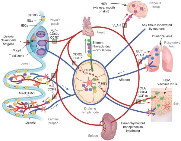
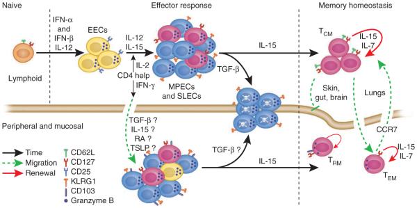
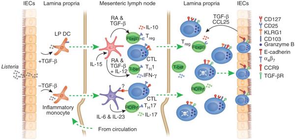
After infection, most antigen-specific memory T cells reside in nonlymphoid tissues. Tissue-specific programming during priming leads to directed migration of T cells to the appropriate tissue, which promotes the development of tissue-resident memory in organs such as intestinal mucosa and skin. Mechanisms that regulate the retention of tissue-resident memory T cells include transforming growth factor-β (TGF-β)-mediated induction of the E-cadherin receptor CD103 and downregulation of the chemokine receptor CCR7. These pathways enhance protection in internal organs, such as the nervous system, and in the barrier tissues--the mucosa and skin. Memory T cells that reside at these surfaces provide a first line of defense against subsequent infection, and defining the factors that regulate their development is critical to understanding organ-based immunity.


Figures

Figure 1
Figure 1
The migration of effector and memory T cells to sites of localized infection. DC acquisition of antigen occurs at peripheral sites such as the lung, skin or gut after a breach in the epithelial layer or local infection. After capturing antigen, DCs access initial afferent lymphatics mediated by signals from integrins, chemokines and semaphorins to migrate to draining lymph nodes to activate naive or memory cells. For example, breach of the intestinal mucosa leads to T cell priming in the MLNs and/or Peyer’s patches. The spleen is poised to generate a substantial T cell response if pathogens are not compartmentally contained and gain access to the bloodstream. In either scenario, a robust T cell population expansion program follows with DC-mediated instruction for migration to the site of initial infection. Activated T cells and circulating memory cells exit the lymph node via the efferent lymphatics and return to circulation through the thoracic duct (colors indicate T cells instructed to home to specific tissues). As T cells migrate through the circulation, integrin and chemokine signals direct their emigration into tissues. In this manner, imprinted T cells have a specific key that allows access to restricted tissues (gates) under normal homeostatic conditions. IEL, intraepithelial lymphocyte; IEC, intestinal epithelial cell; VLA-4, integrin α4β1.
Figure 2
Figure 2
The generation of a diverse and dynamic memory T cell population is a highly orchestrated process. Naive CD8+ T cells rapidly downregulate CD62L and CD127 after exposure to their cognate antigen in the proper contextual environment. IL-12, type I interferons (IFN-α and IFN-β) and IL-2 regulate the initial development of a heterogeneous effector T cell population in lymphoid tissues. CD4+ T cells are thought to be crucial to distinct aspects of this process. After certain infections, such as with l. monocytogenes, CD4+ T cell help is essential for the generation of a robust SLEC population and may also regulate the generation of TCM cells through IL-2 production,. However, IFN-γ produced by CD4+ T cells regulates recruitment of CD8+ T cells to the site of peripheral infection, such after infection of the genital tract with HSV. Once T cells enter peripheral tissues, various molecules from the tissue microenvironment may further influence effector heterogeneity. For example, the cytokines TGF-β and TSLP can have tissue-specific effects on the differentiation of T cells in the intestinal mucosa,,,. Contraction of the T cell response is regulated in part by TGF-β and IL-15, which mediate opposing fates of the effector pool,. At least in lymphoid tissues, TGF-β seems to induce apoptosis of SLECs, whereas IL-15 promotes T cell survival and entry into the memory pool,. TCM cells reside mainly in lymphoid tissues and downregulate expression of granzyme B, whereas TEM and TRM cells reside in peripheral nonlymphoid tissues and maintain granzyme B expression and direct lytic activity. The migration of memory T cells into the skin, gut and brain seems to be restricted, whereas replenishment of liver and lung parenchyma (but not airway) memory T cells occurs through the circulation. Memory T cells located in peripheral tissues may also be regulated by CCR7-mediated emigration signals, which allows the potential for further modification of the TEM cell population,. EEC, early effector cell.
Figure 3
Figure 3
Multifaceted roles of TGF-β in generating mucosal effector and memory T cell populations. TGF-β seems to regulate many aspects of CD8 memory T cell formation in mucosal and potentially other peripheral tissues. Initially, TGF-β derived from intestinal epithelial cells influences the antigen-presenting cells, which migrate to the draining lymph nodes to prime local T cell responses. The presence or absence of TGF-β regulates the expression of CD103 or E-cadherin on antigen-presenting cell subsets in the lamina propria (LP). CD103+ DCs, which also produce TGF-β, mediate imprinting of gut-homing T cells by a mechanism that depends on TGF-β and retinoic acid (RA) and are also responsible for the generation of regulatory T cells (Treg). In conjunction with IL-15, T helper type 1 (TH1) and TH17 cells are ‘preferentially’ generated, which skews the T cell response to a proinflammatory nature. However, TGF-β also suppresses the generation of E-cadherin-positive DCs, which ‘preferentially’ drive a TH17 response in a colitis model. It is unknown whether CD8+ T cells of the SLEC type, which express KLRG1, or other CD103+ CD8+ T cells can interact with E-cadherin expressed by these inflammatory DCs. Whatever holds true, CD4+ T cells primed by these DCs are fully able to migrate to the lamina propria. Once T cells emigrate into the lamina propria, α4β7 is rapidly downregulated and CD103 expression is upregulated in a TGF-β-dependent manner in a subset of lamina propria CD8+ T cells. CCL25 expression by intestinal epithelial cells mediates a chemotactic gradient to recruit effector CD8+ T cells into the epithelium. TGF-β signals also drive the apoptosis of CD8+ T cells of the SLEC type, at least in lymphoid tissues, but may also mediate distinct outcomes on the basis of the presence of other inflammatory or anti-inflammatory mediators,. In this system, TGF-β is probably produced by both the epithelium and lamina propria DCs, but the relative contributions of each source remain unclear. Foxp3, T-bet and RORγt are transcription factors; CTL, cytotoxic T lymphocyte; TGF-βR, TGF-β receptor.

References

    1. Starr TK, Jameson SC, Hogquist KA. Positive and negative selection of T cells. Annu. Rev. Immunol. 2003;21:139–176. - PubMed
    1. Hamann D, et al. Phenotypic and functional separation of memory and effector human CD8+ T cells. J. Exp. Med. 1997;186:1407–1418. - PMC - PubMed
    1. Sallusto F, Lenig D, Forster R, Lipp M, Lanzavecchia A. Two subsets of memory T lymphocytes with distinct homing potentials and effector functions. Nature. 1999;401:708–712. - PubMed
    1. Reinhardt RL, Khoruts A, Merica R, Zell T, Jenkins MK. Visualizing the generation of memory CD4 T cells in the whole body. Nature. 2001;410:101–105. - PubMed
    1. Masopust D, Vezys V, Marzo AL, Lefrançois L. Preferential localization of effector memory cells in nonlymphoid tissue. Science. 2001;291:2413–2417. - PubMed

MeSH terms