. 2011 Dec;17(23-24):3033-44.
doi: 10.1089/ten.TEA.2011.0257. Epub 2011 Aug 29.

An isolated cryptic peptide influences osteogenesis and bone remodeling in an adult mammalian model of digit amputation

Affiliations

An isolated cryptic peptide influences osteogenesis and bone remodeling in an adult mammalian model of digit amputation

Vineet Agrawal et al. Tissue Eng Part A. 2011 Dec.

Abstract

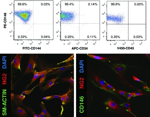
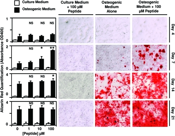
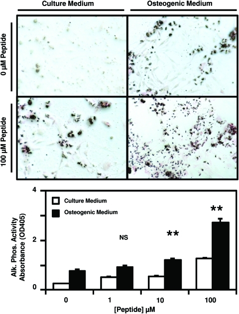
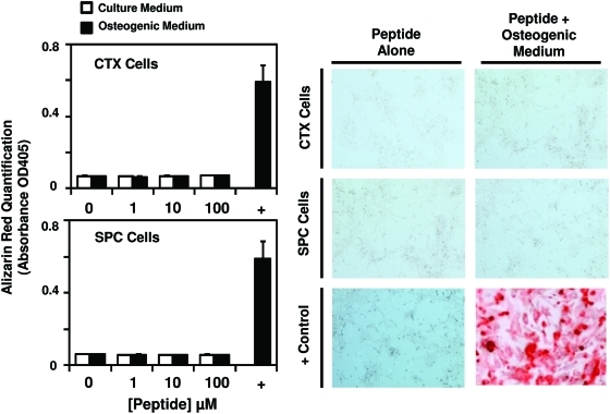
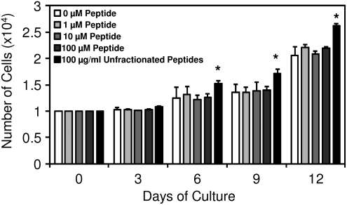
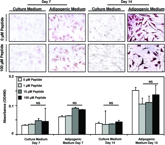
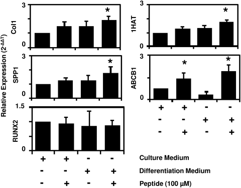
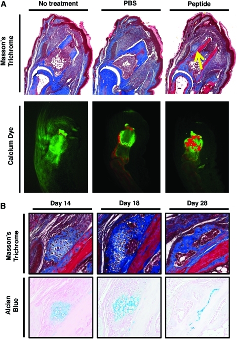
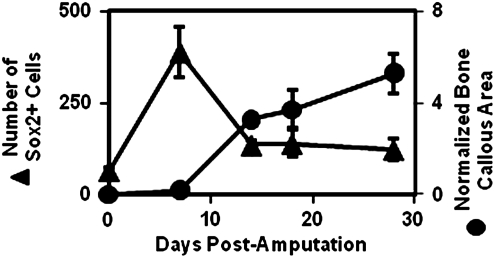
Biologic scaffolds composed of extracellular matrix (ECM) have been used successfully in preclinical models and humans for constructive remodeling of functional, site-appropriate tissue after injury. The mechanisms underlying ECM-mediated constructive remodeling are not completely understood, but scaffold degradation and site-directed recruitment of progenitor cells are thought to play critical roles. Previous studies have identified a cryptic peptide derived from the C-terminal telopeptide of collagen IIIα that has chemotactic activity for progenitor cells. The present study characterized the osteogenic activity of the same peptide in vitro and in vivo in an adult murine model of digit amputation. The present study showed that the cryptic peptide increased calcium deposition, alkaline phosphatase activity, and osteogenic gene expression in human perivascular stem cells in vitro. Treatment with the cryptic peptide in a murine model of mid-second phalanx digit amputation led to the formation of a bone nodule at the site of amputation. In addition to potential therapeutic implications for the treatment of bone injuries and facilitation of reconstructive surgical procedures, cryptic peptides with the ability to alter stem cell recruitment and differentiation at a site of injury may serve as powerful new tools for influencing stem cell fate in the local injury microenvironment.


Figures

FIG. 1.
FIG. 1.
Perivascular stem cells express mesenchymal stem cell markers. Human perivascular stem cells were characterized by flow cytometry and immunostaining to confirm their phenotype as perivascular stem cells. As previously described, perivascular stem cells did not express CD144, CD34, or CD45. They did express perivascular stem cell markers CD146, NG2, and smooth muscle actin. Color images available online at www.liebertonline.com/tea
FIG. 2.
FIG. 2.
Cryptic peptide accelerates osteogenesis of perivascular stem cells. Human perivascular stem cells were cultured in either normal culture medium or osteogenic differentiation medium. After supplementation of medium with 0, 1, 10, or 100 μM of the isolated cryptic peptide, osteogenic differentiation was determined by Alizarin red stain of the cells. At 7 and 14 days postdifferentiation, the isolated cryptic peptide accelerated osteogenesis of perivascular stem cells. *p<0.05, **p<0.01 as compared to the 0 μM osteogenic differentiation group. Error bars represent mean±standard error of the mean (SEM) of experiments in triplicate (n=3). NS, not significant. Color images available online at www.liebertonline.com/tea
FIG. 3.
FIG. 3.
Cryptic peptide increases alkaline phosphatase activity. Human perivascular stem cells were cultured in either culture medium or osteogenic differentiation medium. After supplementation of medium with 0, 1, 10, or 100 μM of the isolated cryptic peptide, alkaline phosphatase activity was measured by p-Nitrophenyl phosphate substrate reaction and staining. At 7 days postdifferentiation and treatment, the isolated cryptic peptide resulted in increased alkaline phosphatase activity. **p<0.01 as compared to the 0 μM osteogenic differentiation group. Error bars represent mean±SEM of experiments in triplicate (n=3). Color images available online at www.liebertonline.com/tea
FIG. 4.
FIG. 4.
Cryptic peptide does not promote osteogenic differentiation in non-osteogenic stem cells. To determine whether the isolated cryptic peptide promotes osteogenic differentiation of nonmesenchymal stem cells, human cortical neuroepithelial stem cells and human spinal cord neural stem cells were cultured in normal culture medium or osteogenic differentiation medium in the presence of 0, 1, 10, or 100 μM of the isolated cryptic peptide. The isolated peptide did not promote osteogenic differentiation of the neural stem cells. Error bars represent mean±SEM of experiments in triplicate (n=3). Color images available online at www.liebertonline.com/tea
FIG. 5.
FIG. 5.
Cryptic peptide does not alter proliferation of perivascular stem cells. To determine whether the peptide induced osteogenesis by increasing proliferation of cells, perivascular stem cells were supplemented in normal growth medium supplemented with 0, 1, 10, or 100 μM peptide, or 100 μg/mL of unfractionated cryptic peptides as a positive control. Over the course of 12 days, no change in cell number was observed after culture in any concentration of cryptic peptide. *p<0.05 as compared to normal growth medium at each time point. Error bars represent mean±SEM of experiments in triplicate (n=3).
FIG. 6.
FIG. 6.
Cryptic peptide does not alter adipogenesis of perivascular stem cells. Human perivascular stem cells were cultured in either culture medium or adipogenic differentiation medium. After supplementation of medium with 0, 1, 10, or 100 μM of the isolated cryptic peptide, differentiation was determined by Oil Red O stain. No differences were noted between treatment groups at any time point. Error bars represent mean±SEM of experiments in triplicate (n=3). Color images available online at www.liebertonline.com/tea
FIG. 7.
FIG. 7.
Cryptic peptide increases expression of osteogenic and chondrogenic genes in vitro. To determine whether the cryptic peptide accelerates osteogenesis by increasing mRNA expression of osteogenic genes, perivascular stem cells were cultured for 4 days in normal growth medium or osteogenic medium unsupplemented or supplemented with 100 μM cryptic peptide. Osteogenic medium supplemented with peptide resulted in a significant increase in Collagen I, Osteopontin (SPP1), 1HAT, and ABCB1 expression. No expression of LPL was observed over 45 cycles of reverse transcriptase-quantitative polymerase chain reaction. *p<0.05 as compared to normal growth medium for each gene. Error bars represent mean±SEM of six experiments (n=6).
FIG. 8.
FIG. 8.
Cryptic peptide promotes bone deposition in an adult mammalian model of digit amputation. To determine whether the isolated cryptic peptide promotes osteogenesis in vivo, adult C57/BL6 mice were subjected to mid-second phalanx amputation and either left untreated, treated with phosphate-buffered saline (PBS) carrier control, or treated with the isolated cryptic peptide. (A) At day 14 postamputation, histologic analysis revealed a bone-like nodule present at the site of amputation in the peptide treated group. Differential calcium dye injections showed that peptide treatment increases calcium deposition at the site of amputation. (B) Alcian blue stain showed that the bone nodule stained positive for glycosaminoglycans at early time points, suggesting that the nodule underwent endochondral ossification. Images are representative of n=4 animals in each treatment group. Color images available online at www.liebertonline.com/tea
FIG. 9.
FIG. 9.
Bone nodule formation correlates with a loss of Sox2+ cells. To determine whether Sox2+ cells may play a role in bone formation, a time course analysis of the accumulation of Sox2+ cells and bone growth, was completed after peptide treatment. After a peak in Sox2+ cell accumulation at 7 days postamputation, a sharp decrease in Sox2+ cells coincided with the histologic appearance and growth of a bone nodule at the site of amputation, consistent with previous studies showing a role for Sox2 in osteogenesis., Error bars represent mean±SEM of experiments in quadruplicate (n=4).

Similar articles

Cited by

References

    1. Caione P. Capozza N. Zavaglia D. Palombaro G. Boldrini R. In vivo bladder regeneration using small intestinal submucosa: experimental study. Pediatr Surg Int. 2006;22:593. - PubMed
    1. Cobb M.A. Badylak S.F. Janas W. Boop F.A. Histology after dural grafting with small intestinal submucosa. Surg Neurol. 1996;46:389. discussion 93–94. - PubMed
    1. Lantz G.C. Badylak S.F. Coffey A.C. Geddes L.A. Blevins W.E. Small intestinal submucosa as a small-diameter arterial graft in the dog. J Invest Surg. 1990;3:217. - PubMed
    1. Hodde J.P. Badylak S.F. Shelbourne K.D. The effect of range of motion on remodeling of small intestinal submucosa (SIS) when used as an achilles tendon repair material in the rabbit. Tissue Eng. 1997;3:27.
    1. Uygun B.E. Soto-Gutierrez A. Yagi H. Izamis M.L. Guzzardi M.A. Shulman C., et al. Organ reengineering through development of a transplantable recellularized liver graft using decellularized liver matrix. Nat Med. 2010;16:814. - PMC - PubMed

Publication types

MeSH terms