

Review
doi: 10.1042/AN20110014.
Current review of in vivo GBM rodent models: emphasis on the CNS-1 tumour model
Affiliations
- PMID: 21740400
- PMCID: PMC3153964
- DOI: 10.1042/AN20110014
Item in Clipboard
Review
Current review of in vivo GBM rodent models: emphasis on the CNS-1 tumour model
ASN Neuro.
.
Abstract
GBM (glioblastoma multiforme) is a highly aggressive brain tumour with very poor prognosis despite multi-modalities of treatment. Furthermore, recent failure of targeted therapy for these tumours highlights the need of appropriate rodent models for preclinical studies. In this review, we highlight the most commonly used rodent models (U251, U86, GL261, C6, 9L and CNS-1) with a focus on the pathological and genetic similarities to the human disease. We end with a comprehensive review of the CNS-1 rodent model.
Figures
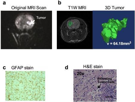
(a) MRI of the original tumour in which the CNS-1 tumour cell line was derived. (b) T1-weighted MRI of 1×10⁵ CNS-1 cells in Lewis rat on day 15. Tumour is highlighted in green. To the right is a 3D reconstruction of the tumour using Mimics Software (Materialise, Leuven, Belgium) by summing the number of voxels that exceeded the intensity threshold in each plane on the T1-weighted subtraction images and multiplying by the appropriate spatial scaling factor (0.35 mm×0.35 mm×1.00 mm per voxel). (c) GFAP staining of the first cell line of CNS-1. (d) An H/E staining of infiltrating tumour cells into normal parenchyma of a Lewis rat brain (scale bar indicates 100 μm).
References
-
- Ali S, Curtin JF, Zirger JM, Xiong W, King GD, Barcia C, Liu C, Puntel M, Goverdhana S, Lowenstein PR, Castro MG. Inflammatory and anti-glioma effects of an adenovirus expressing human soluble Fms-like tyrosine kinase 3 ligand (hsFlt3L): treatment with hsFlt3L inhibits intracranial glioma progression. Mol Ther. 2004;10:1071–1084. - PMC - PubMed
-
- Asai A, Miyagi Y, Sugiyama A, Gamanuma M, Hong SH, Takamoto S, Nomura K, Matsutani M, Takakura K, Kuchino Y. Negative effects of wild-type p53 and s-Myc on cellular growth and tumorigenicity of glioma cells. Implication of the tumor suppressor genes for gene therapy. J Neurooncol. 1994;19:259–268. - PubMed
-
- Auer RN, Del Maestro RF, Anderson R. A simple and reproducible experimental in vivo glioma model. Can J Neurol Sci. 1981;8:325–331. - PubMed
-
- Barth RF. Rat brain tumor models in experimental neuro-oncology: the 9L, C6, T9, F98, RG2 (D74), RT-2 and CNS-1 gliomas. J Neurooncol. 1998;36:91–102. - PubMed
-
- Benda P, Lightbody J, Sato G, Levine L, Sweet W. Differentiated rat glial cell strain in tissue culture. Science. 1968;161:370–371. - PubMed
Publication types
MeSH terms
Grants and funding
LinkOut - more resources
Full Text Sources
Other Literature Sources
Medical
