Review
. 2012 Jun 1;64(8):739-48.
doi: 10.1016/j.addr.2011.06.010. Epub 2011 Jun 29.

Mesenchymal stem cells engineered for cancer therapy

Affiliations
Review

Mesenchymal stem cells engineered for cancer therapy

Khalid Shah. Adv Drug Deliv Rev. .

Abstract

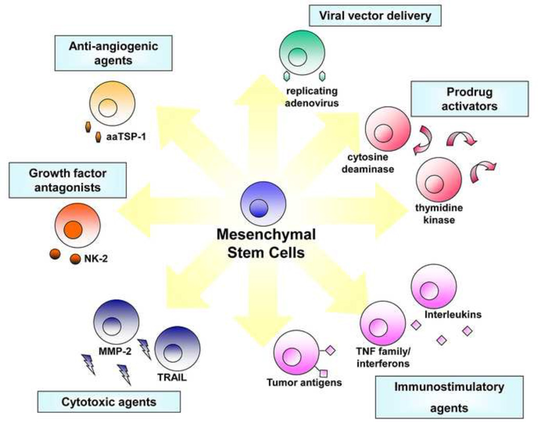
Recent pre-clinical and clinical studies have shown that stem cell-based therapies hold tremendous promise for the treatment of human disease. Mesenchymal stem cells (MSC) are emerging as promising anti-cancer agents which have an enormous potential to be utilized to treat a number of different cancer types. MSC have inherent tumor-trophic migratory properties, which allows them to serve as vehicles for delivering effective, targeted therapy to isolated tumors and metastatic disease. MSC have been readily engineered to express anti-proliferative, pro-apoptotic, anti-angiogenic agents that specifically target different cancer types. Many of these strategies have been validated in a wide range of studies evaluating treatment feasibility or efficacy, as well as establishing methods for real-time monitoring of stem cell migration in vivo for optimal therapy surveillance and accelerated development. This review aims to provide an in depth status of current MSC-based cancer therapies, as well as the prospects for their clinical translation.


Figures

Figure 1
Figure 1. Transgene strategies potentiating MSC for tumor therapy
Tailored to the specific molecular profiles associated with individual tumor types, stem cells can be designed with a variety of different anti-tumor effects.
Figure 2
Figure 2. Migration and Therapeutic efficacy of human mesenchymal stem cells expressing S-TRAIL in mice bearing CSC gliomas
MSC expressing tdTomato were implanted intracranially at a 1 mm distance from established human gliomas expressing GFP-Fluc. (A–B) Photomicrographs showing MSC-tdTomato (red) and gliomas (green) on day 2 (A) and day 10 (B) in brain sections. (C) GBM8 glioma cells were incubated with the conditioned medium from MSC-S-TRAIL and 18 hrs later, GBM8 were analyzed for their viability and casapas-3 activation. Plots showing GBM8 viability incubated with different concentrations of S-TRAIL. (D) Mice bearing established GBM tumors were implanted with MSC-S-TRAIL or control MSC-DsRed2. Survival curves of GBM8-GFP-Fluc-bearing mice treated with MSC-DsRed2 (square) and MSC-S-TRAIL (diamond). (E-J) Photomicrographs showing presence of DsRed2 MSC in brain sections from control mice (E) and Ki67 (F,G) and cleaved caspase-3 (H,I) cells in brain sections from control and MSC-S-TRAIL mice 2 weeks post MSC-implantation. (J) Plot showing the number of cleaved caspase-3 (cells in MSC-S-TRAIL and MSC-DsRed2 treated tumors. Original magnification 10x (B,C); 20X (E-I). Obtained with permission from Proceedings of National Academy of Sciences (PNAS).
Figure 3
Figure 3. Human MSC survive longer in tumor bearing mice and do not influence tumor growth
(A) Fluc bioluminescence intensities of MSC-GFP-Fluc implanted intraparenchymally either alone or mixed with Gli36-EGFRvIII human glioma cells. One representative image of mice with MSC-GFP-Fluc implanted with (+) or without (−) glioma cells is shown. (B–C) Photomicrographs on brain sections from mice 16 days post-implantation showing presence of GFP positive MSC in normal brain (B) and the presence of Ki67 positive glioma cells and GFP positive MSC in glioma bearing brains (C). (D) Fluc bioluminescence intensities of intraparenchymally implanted mice with Gli36-EGFRvIII-FD human glioma cells or a mix of Gli36-EGFRvIII-FD and MSC-GFP. One representative image of mice with Gli36-EGFRvIII-FD implanted with (+) or without (−) MSC-GFP is shown. (E–F) Photomicrographs on brain sections from mice 16 days post-implantation showing expression of DsRed2 in glioma cells (E) and the presence of GFP positive MSC with in mice bearing gliomas (F). Original magnification × 20 (B–C; E–F). Obtained with permission from Proceedings of National Academy of Sciences (PNAS).

References

    1. Jemal A, et al. Cancer statistics, 2007. CA Cancer J Clin. 2007;57(1):43–66. - PubMed
    1. Corsten MF, Shah K. Therapeutic stem-cells for cancer treatment: hopes and hurdles in tactical warfare. Lancet Oncol. 2008;9(4):376–384. - PubMed
    1. Teo AK, Vallier L. Emerging use of stem cells in regenerative medicine. Biochem J. 2010;428(1):11–23. - PubMed
    1. Smith S, Neaves W, Teitelbaum S. Adult stem cell treatments for diseases? Science. 2006;313(5786):439. - PubMed
    1. Momin EN, et al. Mesenchymal stem cells: new approaches for the treatment of neurological diseases. Curr Stem Cell Res Ther. 2010;5(4):326–344. - PubMed

Publication types

Substances