Review
. 2011 Oct;22(10):412-20.
doi: 10.1016/j.tem.2011.05.005. Epub 2011 Jul 7.

Environmental and epigenetic effects upon preimplantation embryo metabolism and development

Affiliations
Review

Environmental and epigenetic effects upon preimplantation embryo metabolism and development

Rebecca J Chason et al. Trends Endocrinol Metab. 2011 Oct.

Abstract

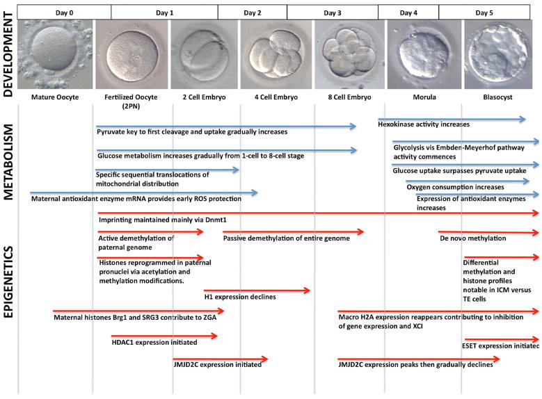
In vitro fertilization has provided a unique window into the metabolic processes that drive embryonic growth and development from a fertilized ovum to a competent blastocyst. Post-fertilization development is dependent upon a dramatic reshuffling of the parental genomes during meiosis, as well as epigenetic changes that provide a new and autonomous set of instructions to guide cellular differentiation both in the embryo and beyond. Although early literature focused simply on the substrates and culture conditions required for progress through embryonic development, more recent insights lead us to suggest that the surrounding environment can alter the epigenome, which can, in turn, impact upon embryonic metabolism and developmental competence.


Figures

Figure 1
Figure 1. Preimplantation Embryo Development, Metabolism and Epigenetic Pathways
Specific timing based on murine model and may differ between species. Development: Preimplantation embryonic development can be categorized into three phases based on gene expression studies [47]. Phase I, from fertilization to the 2-cell stage, is the period of zygotic genome activation (ZGA). Phase II, from the 4-cell to 8-cell stage, is the phase of mid-preimplantation gene activation. Phase III, from the 8-cell to blastocyst stage, is the period of cell differentiation that most notably produces the inner cell mass (ICM) and trophectoderm (TE) cell lineages. Metabolism: Pyruvate is the favored substrate in Phases I and II, with glucose uptake increasing gradually and surpassing pyruvate during Phase III, concomitant with increase in activity of glycolytic pathways. Normal zygote formation (2PN) is accompanied by specific sequential changes in mitochondrial distribution. Oxygen consumption, a viable marker for metabolic activity, increases significantly in Phase III. Stores of maternal mRNA encoding antioxidant enzymes provides ROS protection until ZGA, while embryonic expression becomes notable by blastocyst stage [20]. Epigenetics: DNA methyltransferase 1 (Dnmt1) targets hemi-methylated DNA and maintains imprinting marks during DNA replication. Paternal genome is actively demethylated followed by passive demethylation of maternal genome. De novo methylation begins in the morula and coincides with commencement of cell differentiation. Sperm nuclear protamines are sequentially replaced by acetylated histones then monomethylated histones then di- and tri- methylated histones (H3K4me3, H3K9me2, H3K27me2/3) [47]. Meanwhile, maternal pronuclear histones are modified via acetylation and methylation concomitantly. Linker histone protein H1 declines in cleavage stages likely due to ZGA, while expression of macroH2A, a constituent of the inactive X chromosome heterochromatin, increases in late preimplantation development [47]. Histone deacetylase 1 (HDAC1) appears after fertilization and is vital to preimplantation development [94]. Expression of ERG-associated protein with SET domain (ESET), a histone methyltransferase enzyme, represses CDX2 and is vital to pluripotency of the ICM [95]. JmJD2C, a histone demethylase required for embryo development, appears at 2-cell stage and peaks at 8-cell stage and is involved in NANOG regulation [96].

Similar articles

Cited by

References

    1. Zernicka-Goetz M, et al. Making a firm decision: multifaceted regulation of cell fate in the early mouse embryo. Nat Rev Genet. 2009;10:467–477. - PubMed
    1. Barnett DK, Bavister BD. What is the relationship between the metabolism of preimplantation embryos and their developmental competence? Mol Reprod Dev. 1996;43:105–133. - PubMed
    1. Barnett DK, et al. Glucose and phosphate toxicity in hamster preimplantation embryos involves disruption of cellular organization, including distribution of active mitochondria. Mol Reprod Dev. 1997;48:227–237. - PubMed
    1. Leese HJ, et al. Female reproductive tract fluids: composition, mechanism of formation and potential role in the developmental origins of health and disease. Reprod Fertil Dev. 2008;20:1–8. - PubMed
    1. Hammond J., Jr Recovery and culture of tubal mouse ova. Nature. 1949;163:28. - PubMed

Publication types