

Cytokine expression and microglial activation in progressive supranuclear palsy
- PMID: 21741294
- PMCID: PMC3196843
- DOI: 10.1016/j.parkreldis.2011.06.007
Cytokine expression and microglial activation in progressive supranuclear palsy
Abstract
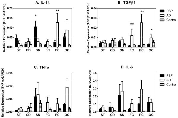
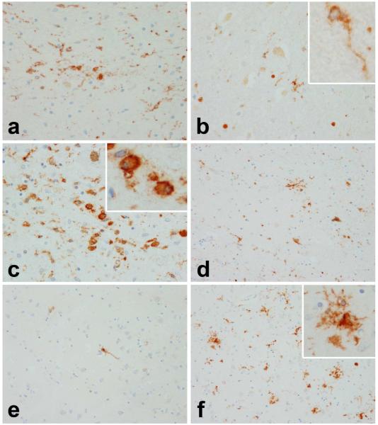
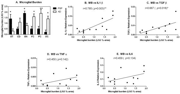
Although little is known about the etiology of progressive supranuclear palsy (PSP), genetic and epigenetic factors, oxidative injury and inflammation are thought to contribute to its development and/or progression. Evidence for activated glia involvement in PSP has raised the possibility that neuroinflammation may contribute to its pathogenesis. To investigate the correlation between neuroinflammation and PSP, a comparative study was conducted on the patterns of cytokine expression in different regions of the brains of PSP, Alzheimer's disease (AD) patients and normal controls. Our results show different patterns of cytokine expression in each disease, with the expression of IL-1β transcripts being significantly higher in the substantia nigra of PSP than in AD and controls, while AD brains had significantly higher IL-1β expression in the parietal cortex compared to PSP and controls. In addition, expression of TGFβ was significantly higher in the cortical areas (particularly frontal and parietal lobes) of AD compared to PSP and controls. These results show a disease-specific topographical relationship among the expression of certain cytokines (IL-1β and TGFβ), microglial activation and neurodegenerative changes, suggesting that these cytokines may contribute to the pathologic process. If so, the use of cytokine-inhibitors and/or other anti-inflammatory agents may be able to slow disease progression in PSP.
Copyright © 2011 Elsevier Ltd. All rights reserved.
Figures
References
-
- Litvan I. Update on progressive supranuclear palsy. Curr Neurol Neurosci Reports. 2004;4:296–302. - PubMed
-
- Williams DR, Lees AJ. Progressive supranuclear plasy: clinicopathological concepts and diagnostic challenges. Lancet Neurol. 2009;8:270–279. - PubMed
-
- Schrag A, Ben-Shlomo Y, Quinn NP. Prevalence of progressive supranuclear palsy and multiple system atrophy: a cross-sectional study. Lancet. 1999;354:1771–5. - PubMed
-
- Hauw JJ, Daniel SE, Dickson D, Horoupian DS, Jellinger K, Lantos PL, et al. Preliminary NINDS neuropathologic criteria for Steele-Richardson-Olszewski syndrome (progressive supranuclear palsy) Neurology. 1994;44:2015–9. - PubMed
-
- Ishizawa K, Dickson DW. Microglial activation parallels system degeneration in progressive supranuclear palsy and corticobasal degeneration. J Neuropathol Exp Neurol. 2001;60:647–57. - PubMed
MeSH terms
Substances
Grants and funding
LinkOut - more resources
Full Text Sources
Other Literature Sources
Medical
Miscellaneous
