. 2011 Oct;64(4):381-92.
doi: 10.1016/j.phrs.2011.06.018. Epub 2011 Jun 29.

Ranolazine reduces Ca2+ overload and oxidative stress and improves mitochondrial integrity to protect against ischemia reperfusion injury in isolated hearts

Affiliations

Ranolazine reduces Ca2+ overload and oxidative stress and improves mitochondrial integrity to protect against ischemia reperfusion injury in isolated hearts

Mohammed Aldakkak et al. Pharmacol Res. 2011 Oct.

Abstract

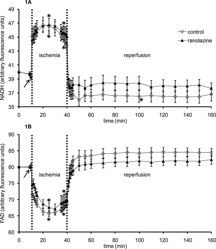
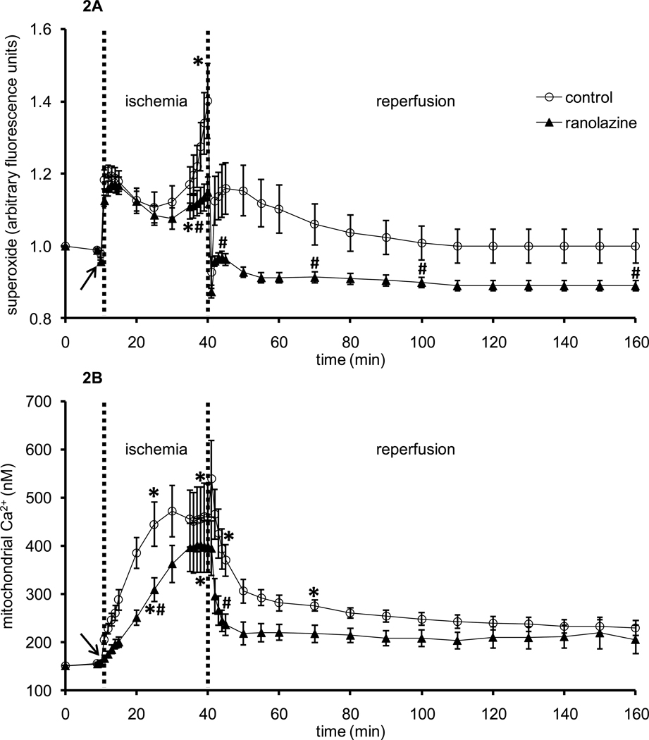
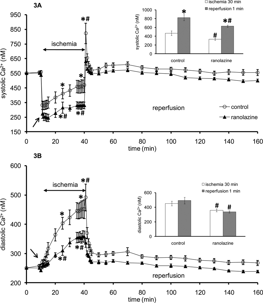
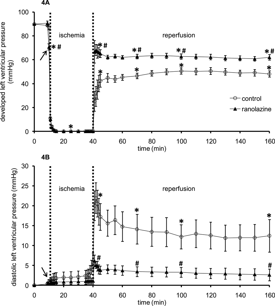
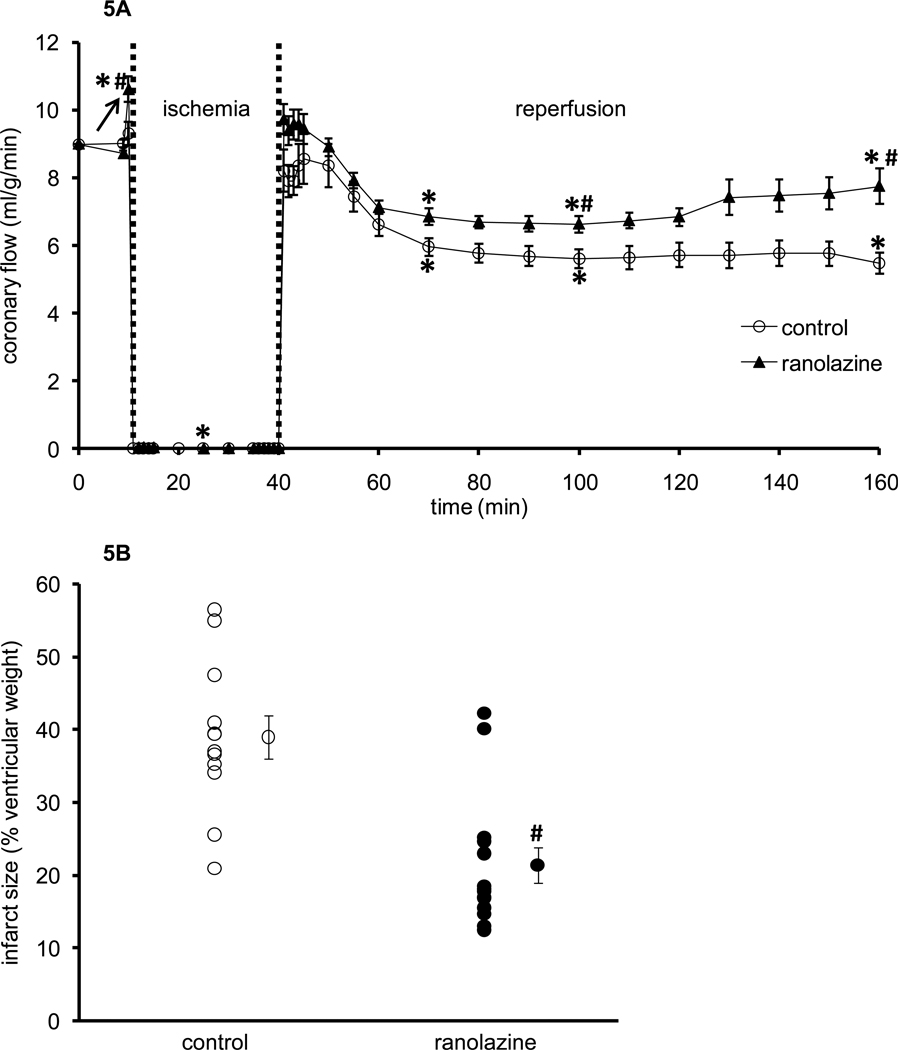
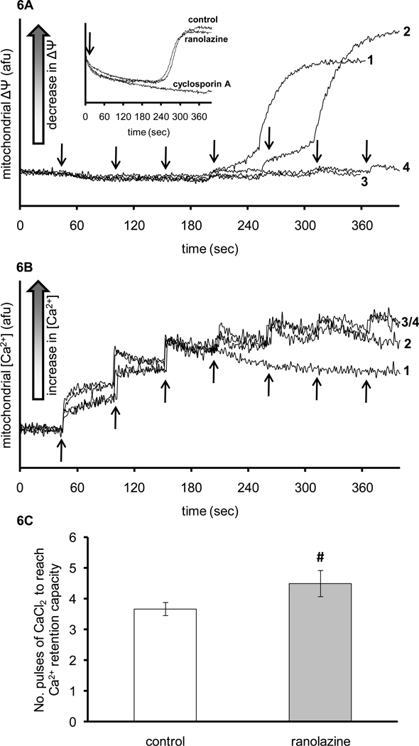
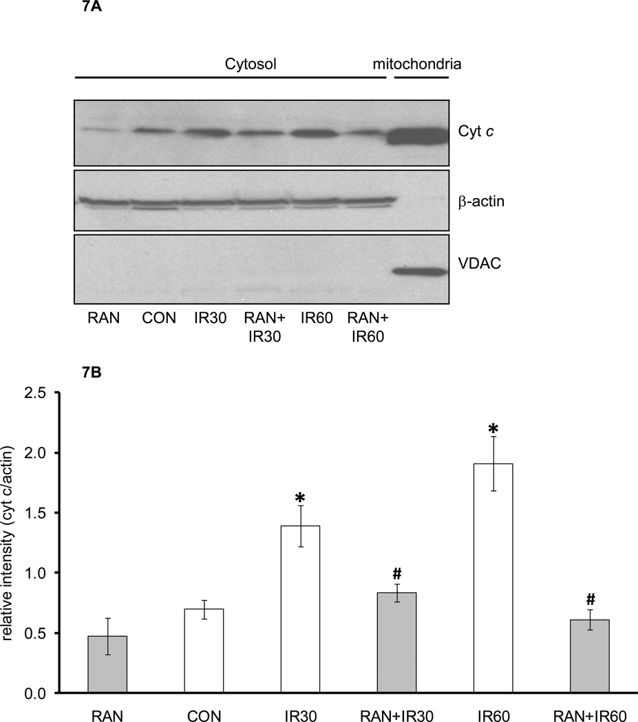
Ranolazine is a clinically approved drug for treating cardiac ventricular dysrhythmias and angina. Its mechanism(s) of protection is not clearly understood but evidence points to blocking the late Na+ current that arises during ischemia, blocking mitochondrial complex I activity, or modulating mitochondrial metabolism. Here we tested the effect of ranolazine treatment before ischemia at the mitochondrial level in intact isolated hearts and in mitochondria isolated from hearts at different times of reperfusion. Left ventricular (LV) pressure (LVP), coronary flow (CF), and O2 metabolism were measured in guinea pig isolated hearts perfused with Krebs-Ringer's solution; mitochondrial (m) superoxide (O2·-), Ca2+, NADH/FAD (redox state), and cytosolic (c) Ca2+ were assessed on-line in the LV free wall by fluorescence spectrophotometry. Ranolazine (5 μM), infused for 1 min just before 30 min of global ischemia, itself did not change O2·-, cCa2+, mCa2+ or redox state. During late ischemia and reperfusion (IR) O2·- emission and m[Ca2+] increased less in the ranolazine group vs. the control group. Ranolazine decreased c[Ca2+] only during ischemia while NADH and FAD were not different during IR in the ranolazine vs. control groups. Throughout reperfusion LVP and CF were higher, and ventricular fibrillation was less frequent. Infarct size was smaller in the ranolazine group than in the control group. Mitochondria isolated from ranolazine-treated hearts had mild resistance to permeability transition pore (mPTP) opening and less cytochrome c release than control hearts. Ranolazine may provide functional protection of the heart during IR injury by reducing cCa2+ and mCa2+ loading secondary to its effect to block the late Na+ current. Subsequently it indirectly reduces O2·- emission, preserves bioenergetics, delays mPTP opening, and restricts loss of cytochrome c, thereby reducing necrosis and apoptosis.


Conflict of interest statement

Conflict of interest: None.

Figures

Figure 1
Figure 1
Changes in NADH autofluorescence (A) and FAD autofluorescence (B) during 1 min treatment with or without ranolazine, and during and after 30 min of no flow global ischemia. Arrow indicates 1 min ranolazine (n=6) or control (n=6) perfusion immediately before ischemia. *P < 0.05, 1 min perfusion before ischemia, during ischemia and reperfusion vs. baseline values; #P < 0.05 ranolazine vs. control.
Figure 2
Figure 2
Changes in DHE fluorescence (indicative of superoxide, O2•−) (A) and indo 1 fluorescence (indicative of m[Ca2+] (B) during 1 min treatment with or without ranolazine, and during and after 30 min of no flow global ischemia. Arrow indicates 1 min ranolazine (n=8 for superoxide; n=6 for m[Ca2+]) or control (n=5 for superoxide; n=6 for m[Ca2+]) perfusion immediately before ischemia. *P < 0.05, 1 min perfusion before ischemia, during ischemia and reperfusion vs. baseline values; #P < 0.05 ranolazine vs. control.
Figure 3
Figure 3
Changes in systolic (A) and diastolic (B) indo 1 fluorescence (indicative of systolic and diastolic [Ca2+], respectively) during 1 min treatment with or without ranolazine, and during and after 30 min of no flow global ischemia. During early ischemia Ca2+ transients continue to occur; once these transients cease, systolic and diastolic Ca2+ are the same. Arrow indicates 1 min ranolazine (n=5) or control (n=7) perfusion immediately before ischemia. *P < 0.05, 1 min perfusion before ischemia, during ischemia and reperfusion vs. baseline values; #P < 0.05 ranolazine vs. control. Insets show systolic (A) and diastolic (B) [Ca2+] at 30 min ischemia (white columns) and at 1 min reperfusion (dark columns). *P < 0.05 [Ca2+] at 1 min reperfusion vs. 30 min ischemia. #P < 0.05 ranolazine vs. control.
Figure 4
Figure 4
Changes in developed left ventricular pressure (A) and diastolic left ventricular pressure (B) during 1 min treatment with or without ranolazine, and during and after 30 min of no flow global ischemia. Arrow indicates 1 min ranolazine (n=14) or control (n=11) perfusion immediately before ischemia. *P < 0.05, 1 min perfusion before ischemia, during ischemia and reperfusion vs. baseline values; #P < 0.05 ranolazine vs. control.
Figure 5
Figure 5
Changes in coronary flow (A) during 1 min treatment with or without ranolazine, and during and after 30 min of no flow global ischemia. Arrow indicates 1 min ranolazine (n=14) or control (n=11) perfusion immediately before ischemia. Panel B shows infarct size as a percentage of total ventricular weight measured after 120 min reperfusion for ranolazine (n=14) and control groups (n=11). *P < 0.05, 1 min perfusion before ischemia, during ischemia and reperfusion vs. baseline values; #P < 0.05 ranolazine vs. control.
Figure 6
Figure 6
Representative traces of rhodamine 123 fluorescence (indicative of membrane potential, ΔΨm) (A) and indo 1 fluorescence (indicative of m[Ca2+]) (B) in mitochondria isolated from IR only hearts (traces 1, 3; n=3) and ranolazine + IR hearts (traces 2, 4; n=3). Mitochondria (0.5 mg/ml) were suspended in KCl-based experimental buffer containing 10 mM pyruvate. In traces 3 and 4, the mitochondrial suspension also contained 0.5 µM cyclosporine A to prevent mPTP opening. The arrows indicate when pulses of 25 µM of CaCl2 were added to the mitochondrial suspension. Panel C shows a summary of the number of CaCl2 pulses needed to reach maximal Ca2+ retention capacity and to initiate ΔΨm depolarization. #P < 0.05 ranolazine vs. control. Inset in panel A represents traces of rhodamine 123 fluorescence (membrane potential, ΔΨm) with 25 µM/min of CaCl2 constantly infused in the mitochondrial suspension with the addition of either ranolazine, cyclosporine A or vehicle as control. Arrow in the inset indicates when ranolazine, cyclosporine A, or vehicle was added.
Figure 7
Figure 7
Representative cytochrome c measurements (A) using Western blotting in cytosolic and mitochondrial fractions. Data are summarized in panel B. Abbreviations are: RAN, hearts treated with ranolazine only without ischemia; CON, hearts not treated with ranolazine and not exposed to ischemia; IR30, hearts exposed to 30 min ischemia and 30 min reperfusion without treatment with ranolazine; RAN+IR30, hearts treated with ranolazine followed by 30 min ischemia and 30 min reperfusion; IR60, hearts exposed to 30 min ischemia followed by 60 min reperfusion without treatment with ranolazine; RAN+IR60, hearts treated with ranolazine followed by 30 min ischemia and 60 min reperfusion. *P < 0.05 ischemia groups vs. no ischemia groups. #P < 0.05 RAN+IR30 or RAN+IR60 vs. IR30 or IR60.

Similar articles

Cited by

References

    1. Yellon DM, Hausenloy DJ. Myocardial reperfusion injury. N Engl J Med. 2007;357:1121–1135. - PubMed
    1. Jennings RB, Sommers HM, Smyth GA, Flack HA, Linn H. Myocardial necrosis induced by temporary occlusion of a coronary artery in the dog. Arch Pathol. 1960;70:68–78. - PubMed
    1. Mewton N, Ivanes F, Cour M, Ovize M. Postconditioning: from experimental proof to clinical concept. Dis Model Mech. 2010;3:39–44. - PubMed
    1. Chen Q, Camara AK, Stowe DF, Hoppel CL, Lesnefsky EJ. Modulation of electron transport protects cardiac mitochondria and decreases myocardial injury during ischemia and reperfusion. Am J Physiol Cell Physiol. 2007;292:C137–C147. - PubMed
    1. Becker LB, vanden Hoek TL, Shao ZH, Li CQ, Schumacker PT. Generation of superoxide in cardiomyocytes during ischemia before reperfusion. Am J Physiol. 1999;277:H2240–H2246. - PubMed

Publication types

MeSH terms