. 2011 Sep;179(3):1104-9.
doi: 10.1016/j.ajpath.2011.05.044. Epub 2011 Jul 8.

In the eye of experimental cerebral malaria

Affiliations

In the eye of experimental cerebral malaria

Raman Saggu et al. Am J Pathol. 2011 Sep.

Abstract

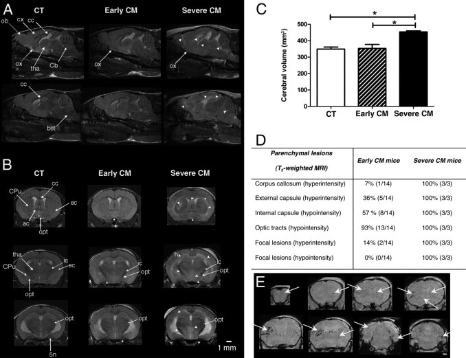
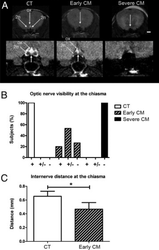
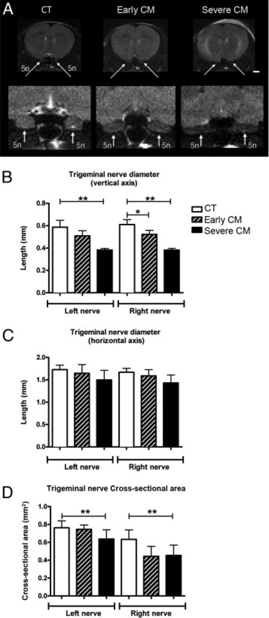
Cerebral malaria is the most severe complication of Plasmodium falciparum infection, accounting for 1 million deaths per year. We characterized the murine disease using in vivo magnetic resonance imaging (MRI) at 4.7 T, proving that ischemic edema is responsible for fatality. The aim of the present study was to identify early markers of experimental cerebral malaria using very high field conventional MRI (11.75 T). CBA/J mice infected with Plasmodium berghei ANKA were observed at an early stage of the disease, before the onset of detectable brain swelling and at the most acute stage of cerebral malaria. Herein, we report the first detection of damage to the optic and trigeminal nerves on T(2)-weighted MRI. The trigeminal nerves appeared hypointense, with significantly reduced diameter and cross-sectional area. The optic nerves were hypointense and often not visible. In addition, the internerve distance between the optic nerves was significantly and progressively reduced between the early and severest stages. Cranial nerve injury was the earliest anatomic hallmark of the disease, visible before brain edema became detectable. Thus, cranial nerve damage may manifest in neurologic signs, which may assist in the early recognition of cerebral malaria.


Figures

Figure 1
Figure 1
Typical brain T2-weighted images of CT and CM mice. A: Sagittal T2-weighted images of CT, early CM, and severe CM brains. Note the brain swelling, crushing of the cerebellum, and hyperintensity of the corpus callosum, indicating edema and demyelination in the severe CM mouse. In addition, focal hyperintensities were detected in the cortex (cx), olfactory bulb (ob), and optic chiasma (ox). The thalamus (tha) exhibited loss of delineation, whereas the hippocampus showed a reduction in contrast. Arrowheads point to changes in T2-contrast. bst, brainstem; Cb, cerebellum; cc, corpus callosum. B: Axial T2-weighted images showing changes in contrast in the corpus callosum (cc), caudate putamen (CPu), external capsule (ec), internal capsule (ic), optic tracts (opt), piriform cortex, and trigeminal nerve (5n) in early CM and severe CM animals compared with the CT mouse. Arrowheads point to changes in T2-contrast. ac, anterior commissure. C: Histogram showing mean cerebral volume of CT, early CM, and severe CM brains calculated from T2-weighted images. Brain volume is significantly increased in severe CM animals compared with in CT and early CM mice (Kruskal-Wallis and Dunn's post hoc tests). Statistical significance for the post hoc test is indicated by *P < 0.05. Error bars indicate SD. D: T2-weighted features of early and severe CM. E: Typical brain T1-weighted images of severe CM mice showing petechiae. Discrete hypointensities (arrows) correspond to focal hemorrhages. Note the extensive lesions in the striatum and cortex. Scale bar = 1 mm.
Figure 2
Figure 2
Morphologic changes of the optic nerve in CM. A: Axial T2-weighted imaging of the optic nerves (2n) in CT, early CM, and severe CM animals. Note the loss of visibility of the chiasmatic region (ox) and the reduction of the internerve distance in early CM mice. Optic nerves at the chiasma level are undetectable in the severe CM mouse. Scale bar = 1 mm. B: Percentage of subjects in which the optic nerve is clearly visible (+), partly visible (+/−), or not visible (−) at the chiasma on T2-weighted images. C: Mean internerve distance between the two optic nerves in CT and early CM animals at the chiasma. Optic nerves are not visible in severe CM mice. The Mann-Whitney U-test showed significantly reduced internerve distance in early CM animals. Error bars indicate SD. Statistical significance is indicated by *P < 0.01.
Figure 3
Figure 3
Morphologic changes of the trigeminal nerve in CM. A: Axial T2-weighted imaging of the trigeminal nerves (5n) in CT, early CM, and severe CM animals. Note the hypointensity and the crushing of the trigeminal nerve in early CM and severe CM mice. Scale bar = 1 mm. B: Histogram representing the mean vertical diameter of the trigeminal nerve in CT and CM mice. Kruskal-Wallis followed by Dunn's post hoc tests showed a significantly reduced diameter in severe CM mice compared with CT animals in both hemispheres and a significant reduction in the right hemisphere in early CM mice. Error bars indicate SD. Statistical significance for the post hoc test is indicated by *P < 0.05, **P < 0.01. C: Histogram representing the mean horizontal diameter of the trigeminal nerve in CT and CM mice. Kruskal-Wallis tests did not reveal any differences in diameter among all the groups. Error bars indicate SD. D: Histogram representing the mean cross-sectional area of the trigeminal nerve. Kruskal-Wallis followed by Dunn's post hoc tests showed a significantly reduced surface area in severe CM compared with CT animals. Error bars indicate 1 SD. Statistical significance for the post hoc test is indicated by **P < 0.01.

Similar articles

Cited by

References

    1. World Health Organization . World Health Organization; Geneva, Switzerland: 2009. World Malaria Report 2009.
    1. Boivin M.J., Bangirana P., Byarugaba J., Opoka R.O., Idro R., Jurek A.M., John C.C. Cognitive impairment after cerebral malaria in children: a prospective study. Pediatrics. 2007;119:e360–e366. - PMC - PubMed
    1. Brewster D.R., Kwiatkowski D., White N.J. Neurological sequelae of cerebral malaria in children. Lancet. 1990;336:1039–1043. - PubMed
    1. Penet M.F., Viola A., Confort-Gouny S., Le Fur Y., Duhamel G., Kober F., Ibarrola D., Izquierdo M., Coltel N., Gharib B., Grau G.E., Cozzone P.J. Imaging experimental cerebral malaria in vivo: significant role of ischemic brain edema. J Neurosci. 2005;25:7352–7358. - PMC - PubMed
    1. Kennan R.P., Machado F.S., Lee S.C., Desruisseaux M.S., Wittner M., Tsuji M., Tanowitz H.B. Reduced cerebral blood flow and N-acetyl aspartate in a murine model of cerebral malaria. Parasitol Res. 2005;96:302–307. - PubMed

Publication types