. 2011 Sep;179(3):1455-70.
doi: 10.1016/j.ajpath.2011.05.031. Epub 2011 Jul 8.

Tumor-recruited neutrophils and neutrophil TIMP-free MMP-9 regulate coordinately the levels of tumor angiogenesis and efficiency of malignant cell intravasation

Affiliations

Tumor-recruited neutrophils and neutrophil TIMP-free MMP-9 regulate coordinately the levels of tumor angiogenesis and efficiency of malignant cell intravasation

Erin M Bekes et al. Am J Pathol. 2011 Sep.

Abstract

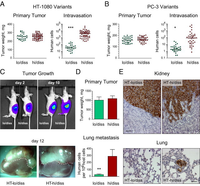
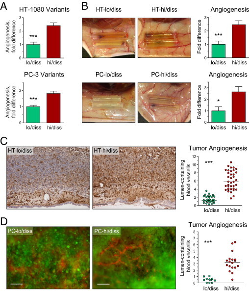
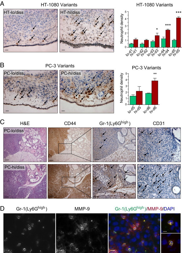
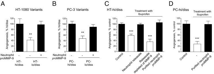
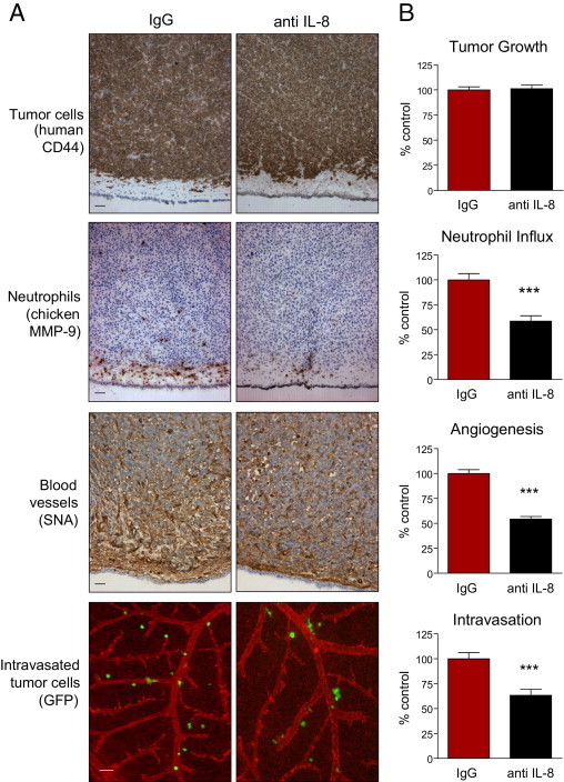
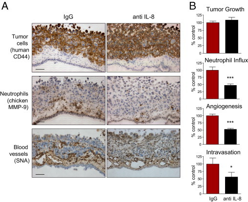
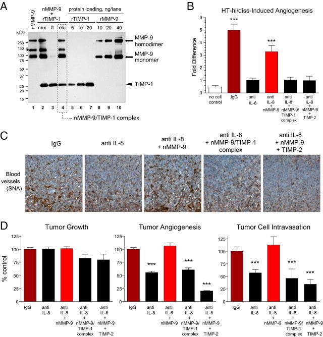
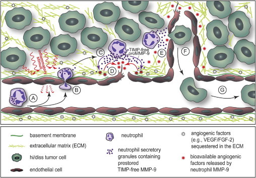
Tumor-associated neutrophils contribute to neovascularization by supplying matrix metalloproteinase-9 (MMP-9), a protease that has been genetically and biochemically linked to induction of angiogenesis. Specific roles of inflammatory neutrophils and their distinct proMMP-9 in the coordinate regulation of tumor angiogenesis and tumor cell dissemination, however, have not been addressed. We demonstrate that the primary tumors formed by highly disseminating variants of human fibrosarcoma and prostate carcinoma recruit elevated levels of infiltrating MMP-9-positive neutrophils and concomitantly exhibit enhanced levels of angiogenesis and intravasation. Specific inhibition of neutrophil influx by interleukin 8 (IL-8) neutralization resulted in the coordinated diminishment of tumor angiogenesis and intravasation, both of which were rescued by purified neutrophil proMMP-9. However, if neutrophil proMMP-9, naturally devoid of tissue inhibitor of metalloproteinases (TIMP), was delivered in complex with TIMP-1 or in a mixture with TIMP-2, the protease failed to rescue the inhibitory effects of anti-IL8 therapy, indicating that the TIMP-free status of proMMP-9 is critical for facilitating tumor angiogenesis and intravasation. Our findings directly link tumor-associated neutrophils and their TIMP-free proMMP-9 with the ability of aggressive tumor cells to induce the formation of new blood vessels that serve as conduits for tumor cell dissemination. Thus, treatment of cancers associated with neutrophil infiltration may benefit from specific targeting of neutrophil MMP-9 at early stages to prevent ensuing tumor angiogenesis and tumor metastasis.


Figures

Figure 1
Figure 1
Dissemination capacity of HT-1080 and PC-3 variants in spontaneous metastasis models. A and B: Tumor growth and intravasation in the chick embryo model. The levels of intravasation of HT-1080 fibrosarcoma (A) or PC-3 prostate carcinoma (B) variants were determined using Alu quantitative PCR in the embryos bearing primary tumors of similar size. Note the logarithmic scale in the intravasation scattergrams. The scattergrams represent cumulative data from nine independent experiments involving grafting of HT-lo/diss (n = 27) and HT-hi/diss (n = 60) and five independent experiments involving grafting of PC-lo/diss (n = 34) and PC-hi/diss (n = 39). ***P < 0.001, two-tailed Student's t-test. C–E: Tumor growth and metastasis of HT-1080 variants in the kidney capsule xenograft model. C: Tumor development of HT-lo/diss and HT-hi/diss variants was analyzed by noninvasive imaging of firefly luciferase-tagged cells using the IVIS system at 2 and 10 days after cell implantation under the renal capsule of immunodeficient mice (top panels). Kidneys with developed tumors were excised from the mice on day 12. Dotted line demarcates the border between the primary tumor and the top of the kidney (bottom panels). D: Efficiency of the primary tumor growth was determined after subtracting the weight of the contralateral kidney. Data from one of six individual experiments involving mice bearing HT-lo/diss (n = 4) and HT-hi/diss (n = 6) tumors (top graph). Levels of lung metastasis in mice bearing HT-lo/diss and HT-hi/diss tumors were quantified using Alu quantitative PCR (bottom graph). Data are given as mean ± SEM from a representative experiment. **P < 0.05, two-tailed Mann-Whitney U-test. E: Immunohistologic detection of human tumor cells within mouse tissues was performed using human-specific CD44 antibody (brown). Top panels: High levels of tumor cell invasion in HT-hi/diss xenografts as compared with their lo/diss counterparts. Scale bar = 50 μm. Bottom panels: Metastatic cells (brown) were detected in the lung from a mouse bearing an HT-hi/diss tumor. Scale bar = 50 μm.
Figure 2
Figure 2
Angiogenesis induced by HT-1080 and PC-3 dissemination variants in avian and mammalian models. A: Angiogenic potential in the chick embryo collagen onplant model. Angiogenic potential of hi/diss variants of HT-1080 fibrosarcoma and PC-3 prostate carcinoma were quantified as fold differences between angiogenic indexes determined in individual onplants compared with their corresponding lo/diss counterparts. Cumulative data from three HT-1080 and five PC-3 independent experiments involving 30 to 79 individual onplants per variant are given as mean ± SEM. *P < 0.05, ***P < 0.001, two-tailed Student's t-test. B: Angiogenic potential in the mouse angiotube model. Panels depict blood vessels converging onto angiotubes. Bar graphs on the right depict fold difference in angiogenesis levels between tumor variants as determined by hemoglobin content in the tubes from three HT-1080 and two PC-3 independent experiments involving 7 to 18 mice per tumor variant. C and D: Tumor angiogenesis in primary CAM tumors. Primary tumors that developed in the chick embryos from HT-1080 variants (C) or PC-3 variants (D) were stained with endothelial cell–specific lectins (SNA or LCA) to visualize blood vessels. Immunohistochemical staining of HT-lo/diss and HT-hi/diss tumors with SNA lectin (C) was followed by quantifying the density of lumen-containing vessels, presented as the scattergram on the right. Scale bar = 100 μm. Number of sections analyzed from three to five individual tumors: HT-lo/diss, n = 30; HT-hi/diss, n = 38. Tumor blood vessels in primary PC-lo/diss and PC-hi/diss tumors were highlighted with red fluorescent-tagged LCA injected into live embryos (D). Scale bar = 100 μm. Quantification of tumor angiogenesis was performed in SNA-stained tumor sections. Number of sections was analyzed from four or five individual tumors: PC-lo/diss, n = 11; PC-hi/diss, n = 18. ***P < 0.001, two-tailed Student's t-test.
Figure 3
Figure 3
Levels of neutrophil influx into primary tumors correlate with tumor cell dissemination potential. A and B: Tumor-recruited neutrophils immunostained with MMP-9–specific antibody (brown) in CAM primary tumors developed from HT-1080 variants (A) and PC-3 variants (B). Arrows point to some of the stained neutrophils. Scale bars = 25 μm. Bar graphs: Density of MMP-9–positive neutrophils in CAM tumors harvested at different times after grafting of HT-1080 and PC-3 dissemination variants. Three to eight individual tumors were analyzed at each time point. *P < 0.05, **P < 0.01, and ***P < 0.001, two-tailed Student's t-test. C: Levels of neutrophil infiltration and angiogenesis in prostate carcinoma orthotopic xenografts correlate with dissemination capacity of lo/diss (upper panels) and hi/diss (bottom panels) tumor variants. Tissue sections from tumors harvested at 4 to 6 weeks after implantation were stained with H&E, human-specific CD44 antibody to discriminate human tumor cells, Gr-1 antibody to detect Ly6G-positive murine neutrophils, and CD31 antibody to highlight blood vessels. Arrows point to some of the Gr-1–positive neutrophils and CD31-positive blood vessels. Scale bars: 200 μm for H&E and CD44 staining; 100 μm for Gr-1 staining; 50 μm for CD31 staining. D: Immunohistologic staining of PC-hi/diss xenografts for Ly6Ghigh with Gr-1 rat mAb and for MMP-9 with rabbit polyclonal antibody. The merged image, also depicting cell nuclei stained with DAPI, indicates that all Gr-1/Ly6Ghigh-positive cells are MMP-9–positive neutrophils. Scale bar = 25 μm. Two merged images on the right depict individual tumor-associated neutrophils at higher magnification of ×630 to illustrate characteristic multilobulated nuclei. Scale bar = 10 μm.
Figure 4
Figure 4
Angiogenic potential of HT-1080 and PC-3 dissemination variants is regulated by neutrophil proMMP-9. A and B: Angiogenesis levels induced by hi/diss and lo/diss variants of HT-1080 fibrosarcoma (A) and PC-3 prostate carcinoma (B) were determined in the CAM collagen onplant model in three to six independent experiments. Low levels of angiogenesis manifested by both types of lo/diss cells were restored by exogenous addition of 2 ng purified neutrophil proMMP-9. Forty-six to 80 individual onplants were analyzed in each group containing fibrosarcoma cells (A), and 20 to 23 individual onplants were analyzed in each group containing prostate carcinoma cells (B). Data are expressed as a percentage of angiogenesis determined in a corresponding hi/diss group and represent mean ± SEM. **P < 0.005, two-tailed Student's t-test. C and D: Levels of angiogenesis diminished by the anti-inflammatory drug ibuprofen were rescued in HT-hi/diss (C) and PC-hi/diss (D) variants by exogenously added neutrophil releasate or 2 ng purified neutrophil proMMP-9 but not by neutrophil releasate specifically depleted of MMP-9 gelatinase by affinity chromatography. Twenty to 94 individual onplants were analyzed for each group containing fibrosarcoma cells (C), and 16 to 22 individual onplants were analyzed in each group containing prostate carcinoma cells (D). Data are expressed as a percentage of angiogenesis determined in the control group (no treatment with ibuprofen and no additives) and represent mean ± SEM. ***P < 0.001, two-tailed Student's t-test.
Figure 5
Figure 5
Specific inhibition of neutrophil influx into HT-hi/diss primary tumors coordinately diminishes tumor angiogenesis and tumor cell intravasation. HT-hi/diss primary tumors were treated with 20 μg control IgG or IL-8/CXCL8-neutralizing antibody (anti–IL-8) in five independent experiments. A: Immunohistochemical analysis of primary tumors. Sections from four to six individual tumors were stained with anti-human CD44 to discriminate human tumor cells (brown), anti-chicken MMP-9 antibody to visualize MMP-9–positive neutrophils (brown), and SNA to highlight blood vessels (brown). Intravasated GFP-tagged HT-hi/diss cells (green) were visualized using live cell imaging in the CAM in which blood vessels were highlighted by red fluorescent LCA. Scale bars = 50 μm. B: Tumor growth, neutrophil influx, tumor angiogenesis, and intravasation were quantified as described in Materials and Methods. Sixty-one and 47 tumor-bearing embryos were analyzed in control IgG- and anti IL-8–treated groups, respectively. Data are expressed as a percentage of IgG control and represent mean ± SEM. ***P < 0.001, two-tailed Student's t-test.
Figure 6
Figure 6
Specific inhibition of neutrophil influx into PC-hi/diss primary tumors coordinately diminishes tumor angiogenesis and tumor cell intravasation. PC-hi/diss primary tumors were treated with 20 μg control IgG or IL-8/CXCL8-neutralizing antibody (anti IL-8) in three independent experiments. A: Immunohistochemical analysis of PC-hi/diss primary tumors. Sections from four to six individual tumors were stained with anti-human CD44 to differentiate human tumor cells (brown), anti-chicken MMP-9 antibody to visualize MMP-9–positive neutrophils (brown), and SNA to highlight blood vessels (brown). Original magnification, ×200. Scale bars = 50 μm. B: Tumor growth, neutrophil influx, tumor angiogenesis, and intravasation were quantified as described in Materials and Methods. Thirty and 22 tumor-bearing embryos were analyzed in control IgG- and anti IL-8–treated groups, respectively. Data are expressed as a percentage of IgG control and represent mean ± SEM. *P < 0.05, and ***P < 0.001, two-tailed Student's t-test.
Figure 7
Figure 7
Rescue of diminished tumor angiogenesis and tumor cell intravasation via delivery of exogenous TIMP-free neutrophil proMMP-9. A: Generation of neutrophil proMMP-9–TIMP-1 complex. Neutrophils isolated from human peripheral blood were induced to release their MMP-9-containing secretory granules, and their proMMP-9 was purified using affinity chromatography (nMMP-9, lane 1). To generate the neutrophil proMMP-9–TIMP-1 complex, purified neutrophil proMMP-9 was mixed with recombinant human TIMP-1 (rTIMP-1) at 1:4 molar ratio (mix, lane 2). After incubation for 1 hour at ambient temperature, the protein mixture (mix) was applied to gelatin Sepharose beads to recover the formed proMMP-9–TIMP-1 complex (elu, lane 4) and remove excess TIMP-1 in the flow-through fraction (ft, lane 3). The proteins were separated using SDS-PAGE under nonreducing conditions, and Western immunoblot analysis was performed using a mixture of mouse anti-human MMP-9 and anti–TIMP-1 mAbs. Lanes 5–7 and 8–10: Loading controls for recombinant TIMP-1 and recombinant proMMP-9, respectively (nanograms per lane). The positions of molecular weight standards are indicated in kilodaltons on the left. Asterisk indicates the position of a 125-kDa heterodimer between proMMP-9 and NGAL (neutrophil gelatinase-associated lipocalin), unique to neutrophils. The molar amounts of proMMP-9 (92 kDa) and TIMP-1 (28 kDa) eluted from gelatin Sepharose beads (dashed box) were calculated via comparison with corresponding protein loading controls and indicated a 1:1 stoichiometric complex between proMMP-9 and TIMP-1. B: Angiogenic and rescuing potential of neutrophil MMP-9 in the chick embryo collagen onplant model. HT-hi/diss cells were incorporated into 3D collagen onplants at 1 × 10⁶ cells/mL with 3 μg/mL normal IgG or IL-8/CXCL8-neutralizing antibody (anti IL-8). Control onplants contained collagen only (no cell control). A subset of the HT-hi/diss–containing collagen onplants treated with anti IL-8 were additionally supplemented with 2 ng purified neutrophil proMMP-9 (nMMP-9) or a stoichiometric 1:1 molar complex between neutrophil proMMP-9 and TIMP1 corresponding to 2 ng nMMP-9 (nMMP-9–TIMP-1) or 2 ng neutrophil proMMP-9 mixed with 5 ng TIMP-2 (eightfold molar excess over nMMP-9). In two independent experiments, 22 to 42 individual onplants were analyzed in each group. Data are expressed as fold difference ± SEM calculated over angiogenesis levels in the anti IL-8 group. ***P < 0.001, two-tailed Student's t-test. C and D: Angiogenic and rescuing capacity of neutrophil MMP-9 in the chick embryo spontaneous metastasis model. HT-hi/diss primary tumors, developing on the CAM, were treated with control IgG or IL-8/CXCL8 neutralizing antibody (anti IL-8) in three independent experiments. A subset of anti-IL-8–treated tumors were additionally supplemented with 30 ng nMMP-9, purified nMMP-9/TIMP-1 stoichiometric complex corresponding to 30 ng nMMP-9, or a mixture of 30 ng nMMP-9 and 70 ng TIMP-2 (7.7-fold molar excess over nMMP-9). C: Immunohistochemical analysis of angiogenesis in primary tumors was performed on tissue sections stained with SNA to highlight blood vessels (brown). Scale bars = 25 μm. D: Tumor growth, angiogenesis, and intravasation were quantified in three independent experiments involving up to 45 tumor-bearing embryos for each treatment condition. Data are expressed as a percentage of IgG control and represent mean ± SEM. ***P < 0.001, two-tailed Student's t-test.
Figure 8
Figure 8
The functional role of neutrophils and their unique TIMP-free MMP-9 in tumor angiogenesis and tumor cell intravasation. Schema demonstrates the attraction of circulating neutrophils to the luminal surface endothelial cells activated by inflammatory stimuli produced by tumor and/or stromal cells (A), followed by neutrophil extravasation into tumor stroma (B), release of secretory granules containing prestored TIMP-free proMMP-9 from induced neutrophils (C), activation of neutrophil proMMP-9 by as yet undefined proteolytic mechanisms to produce the active MMP-9 enzyme catalytically capable of remodeling the ECM and the efficient release of ECM-sequestered angiogenic factors such as VEGF and FGF-2 (D), which in turn induce endothelial cell sprouting and formation of new blood vessels (E), intravasation of tumor cells, likely at specific entry points where ECM and endothelial basement membrane have been proteolytically modified by neutrophil MMP-9 enzyme (F), and dissemination of intravasated tumor cells via circulation (G).

References

    1. Coussens L.M., Werb Z. Inflammation and cancer. Nature. 2002;420:860–867. - PMC - PubMed
    1. Joyce J.A. Therapeutic targeting of the tumor microenvironment. Cancer Cell. 2005;7:513–520. - PubMed
    1. Mantovani A., Allavena P., Sica A., Balkwill F. Cancer-related inflammation. Nature. 2008;454:436–444. - PubMed
    1. Wels J., Kaplan R.N., Rafii S., Lyden D. Migratory neighbors and distant invaders: tumor-associated niche cells. Genes Dev. 2008;22:559–574. - PMC - PubMed
    1. Psaila B., Lyden D. The metastatic niche: adapting the foreign soil. Nat Rev Cancer. 2009;9:285–293. - PMC - PubMed

Publication types

MeSH terms

Substances