. 2011 Jul 13;19(7):930-44.
doi: 10.1016/j.str.2011.05.002.

Protein-RNA and protein-protein recognition by dual KH1/2 domains of the neuronal splicing factor Nova-1

Affiliations

Protein-RNA and protein-protein recognition by dual KH1/2 domains of the neuronal splicing factor Nova-1

Marianna Teplova et al. Structure. .

Abstract

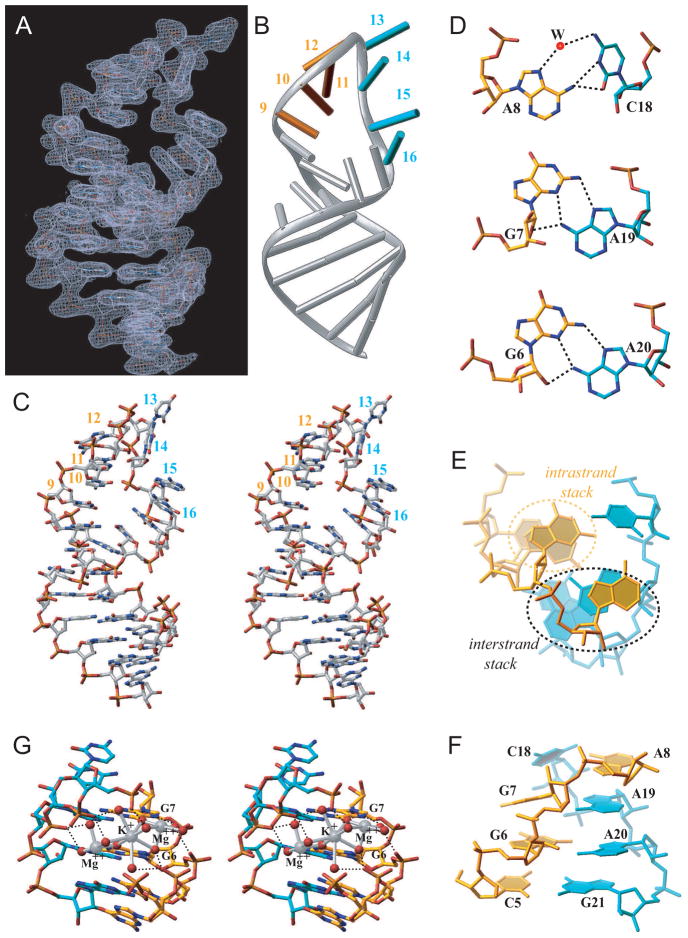
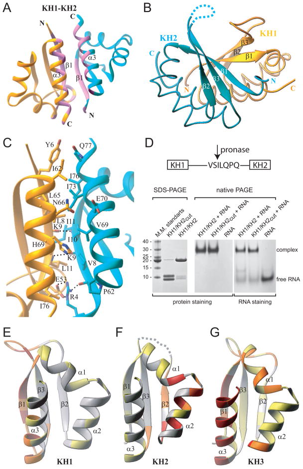
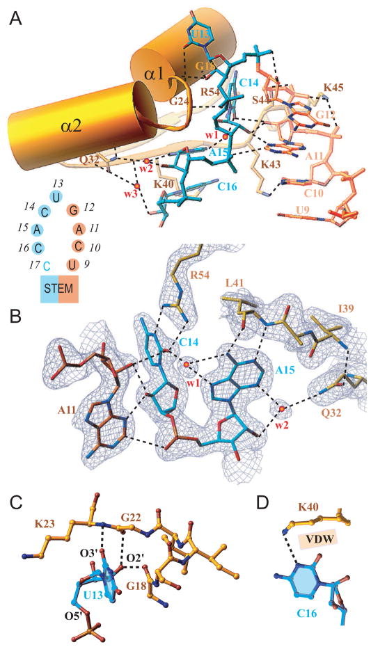
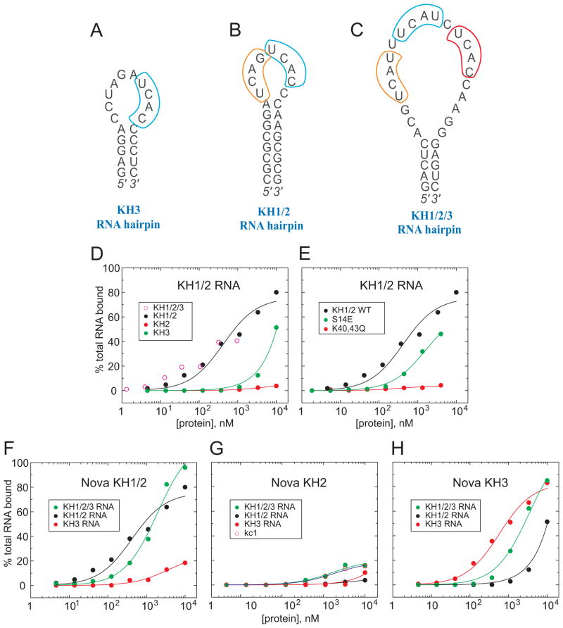
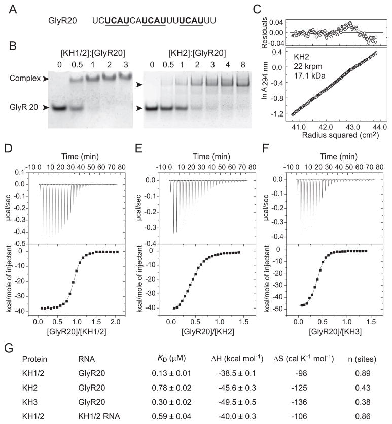
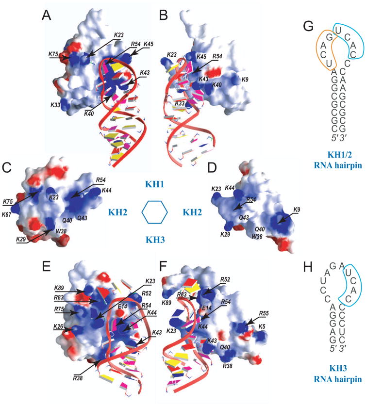
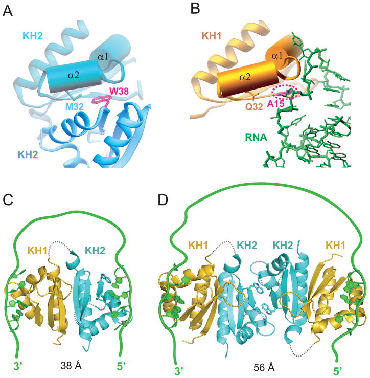
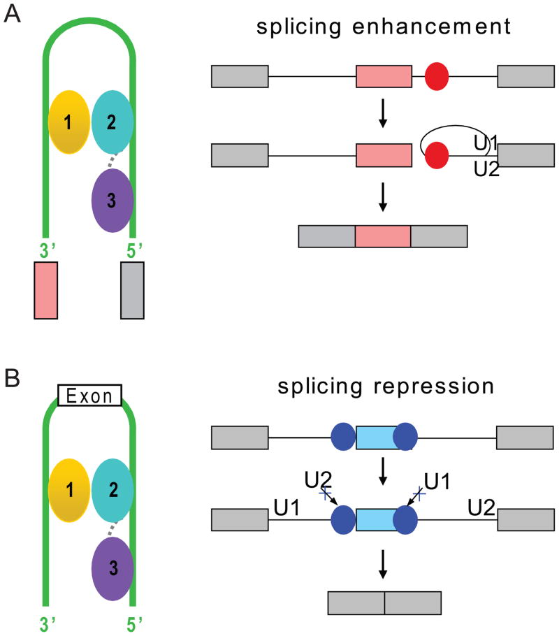
Nova onconeural antigens are neuron-specific RNA-binding proteins implicated in paraneoplastic opsoclonus-myoclonus-ataxia (POMA) syndrome. Nova harbors three K-homology (KH) motifs implicated in alternate splicing regulation of genes involved in inhibitory synaptic transmission. We report the crystal structure of the first two KH domains (KH1/2) of Nova-1 bound to an in vitro selected RNA hairpin, containing a UCAG-UCAC high-affinity binding site. Sequence-specific intermolecular contacts in the complex involve KH1 and the second UCAC repeat, with the RNA scaffold buttressed by interactions between repeats. Whereas the canonical RNA-binding surface of KH2 in the above complex engages in protein-protein interactions in the crystalline state, the individual KH2 domain can sequence-specifically target the UCAC RNA element in solution. The observed antiparallel alignment of KH1 and KH2 domains in the crystal structure of the complex generates a scaffold that could facilitate target pre-mRNA looping on Nova binding, thereby potentially explaining Nova's functional role in splicing regulation.


Figures

Figure 1
Figure 1. Sequences of Nova KH Domains and RNA Hairpin Target and the Structure of the Nova-1 KH1/2-RNA Hairpin Complex in the crystal
(A) Schematic of three KH domains and intervening linkers in Nova proteins. Red-colored numbers indicate the length of linker segments in Nova-1. (B) The Nova-1 KH1/2 construct used in the current project. (C) The sequence of the in vitro selected 25-mer RNA hairpin. The tandem UCAN sites are colored in gold (U9 to G12) and cyan (U13 to C16). Structural studies were also undertaken on a sequence containing a 5BrU2•A24 pair. (D) Sequence and secondary structure (Lewis et al, 1999) alignments of Nova KH1, KH2 and KH3 domains, with conserved residues shown in red. The GXXG motif and so-called variable loop, are denoted as I and V, respectively. (E) Ribbon representation of the structure of the Nova-1 KH1/2-RNA hairpin complex in the crystal. The stoichiometry is KH1/2:RNA hairpin of 2:2. The color codes are RNA in green, KH1 in gold and KH2 in cyan. The UCAC motif interacting with KH1 is colored in blue. See also Table S1, Figures S1, S2 and S3.
Figure 2
Figure 2. RNA Hairpin Architecture, including Hydrogen-bonding, Stacking, and Cation Coordination, in the KH1/2-RNA Hairpin Complex
(A) The RNA hairpin portion of the final |2Fo−Fc| electron density map at 1σ level. (B) The same view of the bound RNA hairpin in a ribbons-and-stick representation. Gold and cyan sticks correspond to U9 to G12 and U13 to C16 segments. (C) Stereo view of the bound RNA hairpin in the complex. Note unusual positioning of one phosphate group (C14-A15 step). (D) Pairing alignments of non-canonical A8•C18, G7•A19 and G6•A20 pairs. (E) Base stack overlap patterns looking down the helical stem axis, highlighting intra-strand and inter-strand overlap patterns. (F) Cross-strand stacking between A8 and A19 within the zippered-up stem in the complex. See also Figure S1. (G) Stereo pair highlighting hydrated Mg2+ and K+ cations positioned within the major groove of the zippered up stem segment. The cations are shown as large silver spheres while the water oxygens and O6 groups of G6 and G7 are shown as small red spheres. Hydrogen bonds are shown as black dashed lines, while ion coordination are shown as silver bonds.
Figure 3
Figure 3. Ribbon Representations of KH1/2 Interface in the KH1/2-RNA Hairpin Complex and Distribution of the Protein-Protein Interaction Propensities over the Surface of Nova KH Domains
(A) A global overview of the KH1(gold)/KH2(cyan) interface, with the segments constituting the interface, colored in pink. (B) The three-stranded β-sheets of the KH1 and KH2 domains form a continuous twisted six-stranded β-sheet. (C) Details of the KH1/2 interface, highlighting interactions between side chains of residues involved in inter-facial contacts. (D) SDS-PAGE electrophoresis (left panel) showing pronase cleavage resulting in two protein fragments (cleavage at Ser90-Ile91 step) with molecular mass of approximately 9 and 10 kDa. Native PAGE (middle and right panels) establishing complex formation between KH1/KH2 and RNA, regardless of cleavage of the linker. (E to G) Distribution of the protein-protein interaction propensities over the protein surface as calculated by the Optimal Docking Area (ODA) approach for three different KH domains. Ribbons representation of KH1 (panel E, present study) and KH2 domains of the Nova-1 KH1/2 protein (panel F, present study) and Nova-2 KH3 domain (panel G, Lewis et al., 2000). Structures are colored by the absolute magnitude of the ODA signal from the strongest in red, through medium in orange and weak in yellow, to the weakest in white. See also Figure S5.
Figure 4
Figure 4. KH1-RNA Interactions in Complex
(A) Overview of interactions between RNA hairpin loop (U9 to G12 in gold and the U13 to C16 in cyan, see insert) and the N-terminal half of the KH1 domain (Tyr15 to Lys45 in gold, encompassing α1, loop I, α2 and β2 segments). Bridging water molecules at the interface are labeled w1, w2 and w3. (B) Details of intermolecular contacts involving the C14-A15 segment of the RNA, and amino acids Arg54, Lys41 and Gln32 of the KH1 domain. Note buttressing interactions between the base and sugar of A11 and C14. The side chain of Lys40 is removed for clarity. (C) Details of intermolecular contacts involving U13 base and sugar hydroxyl group of the RNA, and backbone atoms of amino acids Gly18, Gly22 and Lys23 of the KH1 domain. (D) Intermolecular contacts between the Watson-Crick edge of C16 and the side chain amine (hydrogen bonding) and methylene groups (van der Waals) of Lys40.
Figure 5
Figure 5. Filter Binding Assays for Nova KH Domain-RNA Complexes
(A) The sequence of the Nova-2 KH3-binding 20-nt RNA hairpin containing a single UCAN site (Lewis et al., 2000). (B) The sequence of the Nova-1 KH1/2-binding 25-nt RNA hairpin containing a (UCAN)2 site (Musunuru and Darnell, 2004) that was used in this work. (C) The sequence of the Nova-1 KH1/2/3-binding 32-nt RNA hairpin containing a (UCAN-N)3 site (Buckanovich and Darnell, 1997). (D) Filter-binding assays for complex formation of Nova KH1/2/3 (open circles), KH1/2 (black circles), KH2 (red circles), and KH3 (green circles) with KH1/2 RNA hairpin containing a (UCAN)2 site (panel B). (E) Filter-binding assays for complex formation of wild-type Nova KH1/2 (black circles), Ser14Glu mutant (green circles), and Lys40Gln, Lys43Gln dual mutant (red circles) with KH1/2 RNA hairpin containing a (UCAN)2 site (panel B). (F) Filter-binding assays for complex formation of Nova KH1/2 with RNA hairpins containing a single UCAN site (panel A) (red circles), (UCAN)2 site (panel B) (black circles), and (UCAN-N)3 site (panel C) (green circles). (G) Filter-binding assays for complex formation of Nova KH2 with RNA hairpins containing a single UCAN site (panel A) (red circles), (UCAN)2 site (panel B) (black circles), (UCAN-N)3 site (panel C) (green circles), and a RNA hairpin specific for FMRP KH2 domain (open circles). (H) Filter-binding assays for complex formation of Nova KH3 with RNA hairpins containing a single UCAN site (panel A) (red circles), (UCAN)2 site (panel B) (black circles), and (UCAN-N)3 site (panel C) (green circles).
Figure 6
Figure 6. NMR-monitored Complexation Shifts in Nova-1 KH2 Domain on Addition of UCAC RNA Tetranucleotide
(A) Selected regions of 1H,15N-HSQC of Nova-1_KH2 in presence of various amount of UCAC RNA tetranucleotide (1:0 left; 1:4 middle; 1:36 right). Cross peaks of V28 in free (V28f) and RNA-bound state (V28c) are shown. (B) Histogram outlining the magnitude of the average chemical shift perturbation of the 15N and 1H backbone amide resonances of Nova-1 KH2 on complex formation with UCAC RNA tetranucleotide. The average chemical shift difference Δδave between the free and RNA-bound forms of KH2 was calculated using a correlation: Δδave=ΔδH2+0.1ΔδN2, where ΔδH is the chemical shift of amide proton and ΔδN is the chemical shift of amide nitrogen. Protein secondary structure elements are indicated on the top. (C) Comparison of the residues involved in KH2-UCAC interactions indicated by the average amide chemical shift perturbations (left panel) with the residues involved in direct KH1-UCAC interactions observed in the crystal structure of KH1/2-RNA hairpin complex (right panel). The RNA is shown in green, the KH1 residues directly contacting U13-C14-A15-C16 segment are shown in red on the KH1 domain backbone (right panel), with the magnitude of the KH2 complexation shifts on RNA binding color-coded on the KH2 domain backbone with red representing the largest and grey the smallest shifts (left panel). See also Figure S4.
Figure 7
Figure 7. Binding of Nova-1 tandem KH1/2 and single KH2 and KH3 domains to UCAU repeat element of GlyRα2 pre-mRNA
(A) RNA sequence of an intronic 20-nt UCAU repeat element of GlyRα2 pre-mRNA used in binding experiments. Three non-overlaping UCAU repeats (underlined) separated by 2 nucleotides represent potential binding sites for individual Nova-1 KH domains. (B) Electrophoretic mobility gel shift data for binding of KH1/2 to GlyR20 establishing 1:1 stoichiometry of the complex (left panel); and of KH2 to GlyR20 establishing 2:1 stoichiometry of the complex (right panel). (C) Analytical ultracentrifugation data of a 30 μM sample of KH2 at 20°C and 22,000 rpm in 25 mM Tris-HCl (pH 7.5) and 500 mM KCl. (D–F) Isothermal titration calorimetry (ITC) binding curves of GlyR20 binding to KH1/2 (panel D), KH2 (panel E) and KH3 (panel F) domains. A thermogram as a result of titration is shown in the top panel, and a plot of the total heat released as a function of the molar RNA/protein ratio is shown in the bottom panel. Solid lines indicate non-linear least-squares fit to a theoretical titration curve using Microcal software, with ΔH (binding enthalpy kcal mol−1), KD (association constant), and n (number of binding sites per monomer) as variable parameters. (G) Summary of ITC-based energetic parameters for tandem and single KH-RNA complexes.
Figure 8
Figure 8. Two Perpendicular Views of the Electrostatic Surface of Nova KH1 (with bound RNA), KH2 and KH3 (with bound RNA) Domains
(A, B) Two alternate views of KH1 domain of KH1/2 complexed with RNA hairpin containing tandem UCAN-sites within the loop segment. (C, D) Two alternate views of KH2 domain of KH1/2, that does not form a complex with RNA hairpin. (E, F) Two alternate views of KH3 domain complexed with RNA hairpin containing single UCAC site (Lewis et al., 2000; PDB entry 1EC6). Blue and red patches are associated with positively- and negatively-charged KH surface segments. The RNA backbone is shown in a ribbon and the bases in a slab representation. The slabs are colored as follows: U, blue; C, magenta; A, red; G, yellow. (G) The sequence of the Nova-1 KH1/2-binding 25-nt RNA hairpin containing a (UCAN)2 site (Musunuru and Darnell, 2004) that was used in this work. (H) The sequence of the Nova-2 KH3-binding 20-nt RNA hairpin containing a single UCAN site (Lewis et al., 2000).
Figure 9
Figure 9. Models for Interaction of Nova KH1/2 Domains with Wild-type RNA Targets through a RNA Looping Mechanism
(A) The KH2-KH2 interaction between symmetry-related molecules in the crystal of the KH1/2-RNA hairpin complex. Note the juxtaposition of Met32 and Trp38 within van der Waals contact across the interface. (B) The KH1 (gold) - RNA (green) interface in the KH1/2-RNA hairpin complex. Note the juxtaposition of Gln32 on KH1 and A15 on the RNA within van der Waals contact across the intermolecular interface. (C) Model where Nova-1 KH1/2 monomer (based on ultracentrifugation data in solution) is targeted by a RNA containing a pair of sequence elements capable of targeting KH1 (with high affinity) and KH2 (most likely with lower affinity) and separated by a linker segment of sufficient length. The stoichiometry of this complex is 1:1 Nova-1 KH1/2:RNA. (D) Model of Nova-1 KH1/2 based on the x-ray structure of the complex (Figure 2A), where the RNA-binding surfaces of the KH2 domains pack against each other in the crystal lattice. The target RNA contains a pair of sequence elements capable of targeting KH1 with high affinity and separated by a longer linker segment of sufficient length. The stoichiometry of this complex is 2:1 Nova-1 KH1/2:RNA.
Figure 10
Figure 10. Models of RNA looping induced by Nova KH1–3 domains binding to splicing enhancers and splicing silencers predicted by Nova RNA map of splicing regulation (Ule et al., 2006)
(A) Model based on the examples of Nova upregulation of exon inclusion by binding to intronic splicing enhancer elements (red circles) to enhance spliceosome assembly. (B) Model based on the examples of Nova inhibition of exon inclusion by binding to exonic splicing silencing element (blue circle) and blocking U1 snRNP (U1) assembly on the pre-mRNA, and Nova inhibition of exon inclusion by binding to intronic splicing silencing element immediately upstream of alternative exon by blocking recognition of the 3′ splice site by U2 snRNP (U2). KH1, KH2 and KH3 are colored gold, cyan and purple, respectively. The linkers between KH1 and KH2, and between KH2 and KH3 are represented by the dotted gray lines. The green line represents RNA loop.

Similar articles

Cited by

References

    1. Albert ML, Darnell RB. Paraneoplastic neurological degenerations: keys to tumour immunity. Nat Rev Cancer. 2004;4:36–44. - PubMed
    1. Beuth B, Garcia-Mayoral MF, Taylor IA, Ramos A. Scaffold-independent analysis of RNA-protein interactions: the Nova-1 KH3-RNA complex. J Am Chem Soc. 2007;129:10205–10210. - PubMed
    1. Beuth B, Pennell S, Arnvig KB, Martin SR, Taylor IA. Structure of a Mycobacterium tuberculosis NusA-RNA complex. EMBO J. 2005;24:3576–3587. - PMC - PubMed
    1. Buckanovich RJ, Darnell RB. The neuronal RNA binding protein Nova-1 recognizes specific RNA targets in vitro and in vivo. Mol Cell Biol. 1997;17:3194–3201. - PMC - PubMed
    1. Burd CG, Dreyfuss G. Conserved structures and diversity of functions of RNA-binding proteins. Science. 1994;265:615–621. - PubMed

Publication types

MeSH terms

LinkOut - more resources