. 2011 Jul 13;19(7):1011-20.
doi: 10.1016/j.str.2011.03.023.

Insights into the evolution of a complex virus from the crystal structure of vaccinia virus D13

Affiliations

Insights into the evolution of a complex virus from the crystal structure of vaccinia virus D13

Mohammad W Bahar et al. Structure. .

Abstract

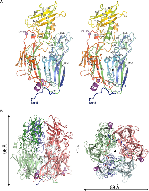
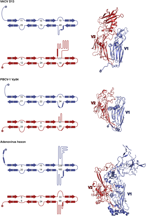
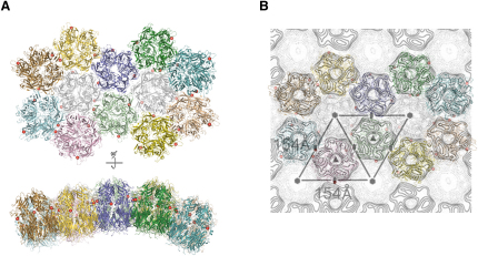
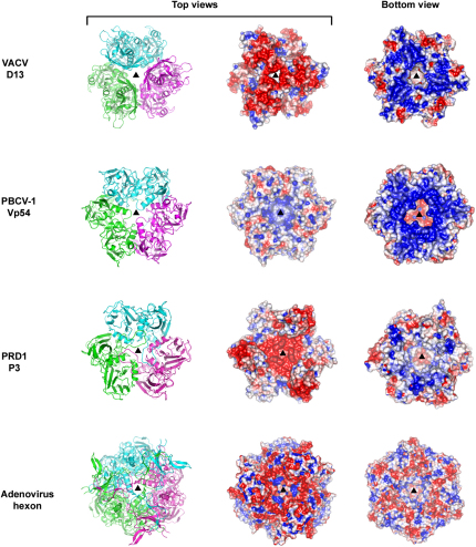
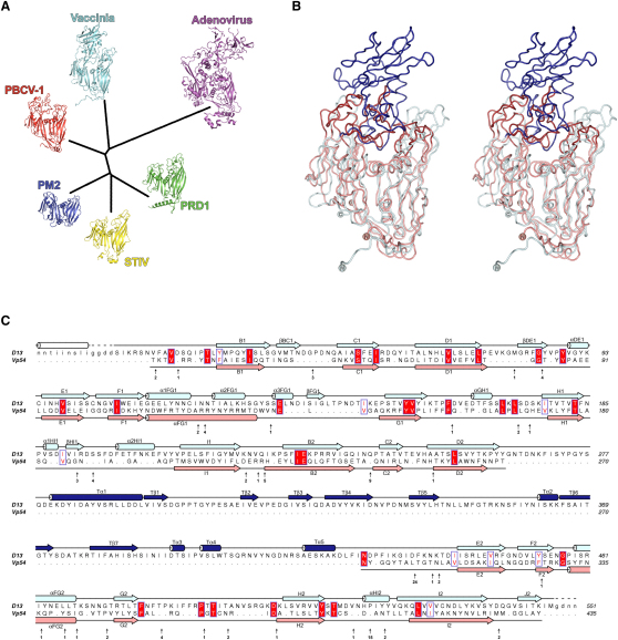
The morphogenesis of poxviruses such as vaccinia virus (VACV) sees the virion shape mature from spherical to brick-shaped. Trimeric capsomers of the VACV D13 protein form a transitory, stabilizing lattice on the surface of the initial spherical immature virus particle. The crystal structure of D13 reveals that this major scaffolding protein comprises a double β barrel "jelly-roll" subunit arranged as pseudo-hexagonal trimers. These structural features are characteristic of the major capsid proteins of a lineage of large icosahedral double-stranded DNA viruses including human adenovirus and the bacteriophages PRD1 and PM2. Structure-based phylogenetic analysis confirms that VACV belongs to this lineage, suggesting that (analogously to higher organism embryogenesis) early poxvirus morphogenesis reflects their evolution from a lineage of viruses sharing a common icosahedral ancestor.


Figures

Figure 1
Figure 1
Structure of VACV D13 (A) Stereo ribbon representation of a monomer of D13 colored from blue (N terminus) to red (C terminus). The turret is treated as a separate domain with α helices and β strands labeled with prefix “T” for clarity. The N-terminal helix present in D13D513G is shown in magenta. The position of the D513G mutation is shown with a sphere, colored magenta, and labeled. (B) Structure of the D13 trimer viewed from the side (left) and from below (right). Each subunit is colored separately, with N-terminal tails and turret domains being a darker shade compared to the jelly-rolls. Approximate molecular dimensions are shown, and the N-terminal helix present in D13D513G is colored magenta. The black triangle denotes the molecular 3-fold symmetry axis. See also Figure S1.
Figure 2
Figure 2
Comparison of VACV D13, PBCV-1 Vp54, and Adenovirus Hexon Capsid Proteins Topology diagrams (left) depict the arrangement of the β strands (arrows) and certain α helices (rectangles) of each jelly-roll. V1 (N-terminal) and V2 (C-terminal) jelly-rolls are colored blue and red respectively. The numbers of residues present in the loops connecting the β strands are indicated. Ribbon diagrams of D13, Vp54, and hexon are shown on the right with the BIDG and CHEF β sheets labeled for each jelly-roll. The N- and C-termini are shown as spheres and labeled. See also Table S1.
Figure 3
Figure 3
Structural Model of Trimer-Trimer Interactions in the D13 Lattice VACV D13 trimers were superposed using SHP (Stuart et al., 1979) onto a facet of P3 capsid proteins from the related dsDNA virus PRD1. (A) The lattice is viewed, in the top, from the membrane looking up with a view perpendicular to the membrane (out of the virus particle), whereas the bottom shows the view tangential to the viral crescent membrane (note the curvature of the IV particle will be somewhat less, because it is larger than PRD1). Individual trimers are colored separately, and the position of the D513G mutation is shown with a red sphere. Two D13 trimers are drawn semitransparent and colored gray, to show the positions of systematic absences of trimers in the VACV IV lattice that give rise to the honeycomb structure. (B) The D13 trimers from (A) are superimposed onto the projection density of the orfv075 lattice II, which is taken to be a model for the honeycomb lattice seen on the IV particles (Figure 4B of Hyun et al., 2007, reproduced with permission from the Journal of Virology).
Figure 4
Figure 4
Comparison of Surface Charge Distribution on dsDNA Virus MCPs Trimers of D13, Vp54, P3, and hexon are shown as cartoons (left, each subunit colored separately) and electrostatic surfaces (middle and right) and viewed from the top and bottom along their 3-fold symmetry axes (black triangles). Electrostatic charges are contoured from red (−3 kT/e) to blue (+3 kT/e).
Figure 5
Figure 5
Structure-Based Phylogenetic Tree of the PRD1-Adenovirus Lineage and Comparison of VACV D13 to PBCV-1 Vp54 (A) Structures of capsid proteins from vaccinia (D13), PBCV-1 (Nandhagopal et al., 2002), adenovirus (Rux et al., 2003), STIV (Khayat et al., 2005), PRD1 (Benson et al., 2002), and PM2 (Abrescia et al., 2008) were superposed and a pairwise distance matrix was constructed as described previously (Bamford et al., 2005; Riffel et al., 2002). Cartoons of each capsid protein are drawn at the branch ends, colored separately and labeled with their virus names. (B) Stereo view of VACV D13 and PBCV-1 Vp54 superposed by their Cα traces, with their core jelly-roll domains colored light blue and salmon, and the turret domain and tower loops dark blue and red, respectively. N- and C-termini are shown as spheres and labeled. (C) Structure-based sequence alignment of VACV D13 and PBCV-1 Vp54 derived from the superposition via SHP (Stuart et al., 1979). The secondary structures of D13 and Vp54 are drawn above and below the alignment respectively, and colored as in (B); β strands are shown as arrows and α helices as cylinders. The N-terminal helix seen in D13D513G is drawn as a white cylinder. Strictly conserved residues are boxed in red, moderately conserved residues are boxed white with a red face, and conservation is scored according to the Blosum62 scoring matrix. Residues of Vp54 that are not aligned by the structural superposition are omitted and their number and positions are indicated under the Vp54 sequence with black arrows. See also Table S1.

Comment in

  • Pass the jelly rolls.
    Khayat R, Johnson JE. Khayat R, et al. Structure. 2011 Jul 13;19(7):904-6. doi: 10.1016/j.str.2011.06.004. Structure. 2011. PMID: 21742257 Free PMC article.

Similar articles

Cited by

References

    1. Abrahams J.P., Leslie A.G. Methods used in the structure determination of bovine mitochondrial F1 ATPase. Acta Crystallogr. D Biol. Crystallogr. 1996;52:30–42. - PubMed
    1. Abrescia N.G., Cockburn J.J., Grimes J.M., Sutton G.C., Diprose J.M., Butcher S.J., Fuller S.D., San Martin C., Burnett R.M., Stuart D.I. Insights into assembly from structural analysis of bacteriophage PRD1. Nature. 2004;432:68–74. - PubMed
    1. Abrescia N.G., Grimes J.M., Kivela H.M., Assenberg R., Sutton G.C., Butcher S.J., Bamford J.K., Bamford D.H., Stuart D.I. Insights into virus evolution and membrane biogenesis from the structure of the marine lipid-containing bacteriophage PM2. Mol. Cell. 2008;31:749–761. - PubMed
    1. Afonine, P.V., Grosse-Kunstleve, R.W., and Adams, P.D. (2005). CCP4 Newsletter 42, contribution 8.
    1. Athappilly F.K., Murali R., Rux J.J., Cai Z., Burnett R.M. The refined crystal structure of hexon, the major coat protein of adenovirus type 2, at 2.9 A resolution. J. Mol. Biol. 1994;242:430–455. - PubMed

Publication types

MeSH terms

Substances

LinkOut - more resources