. 2011 Sep 1;417(2):281-92.
doi: 10.1016/j.virol.2011.05.015. Epub 2011 Jul 13.

Osteopontin upregulation in rotavirus-induced murine biliary atresia requires replicating virus but is not necessary for development of biliary atresia

Affiliations

Osteopontin upregulation in rotavirus-induced murine biliary atresia requires replicating virus but is not necessary for development of biliary atresia

Paula M Hertel et al. Virology. .

Abstract

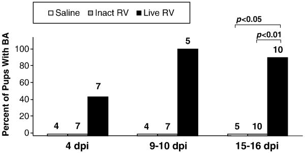
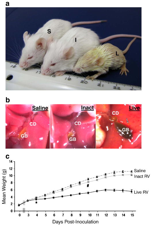
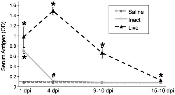
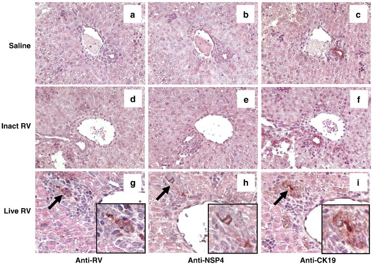
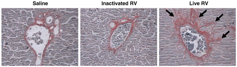
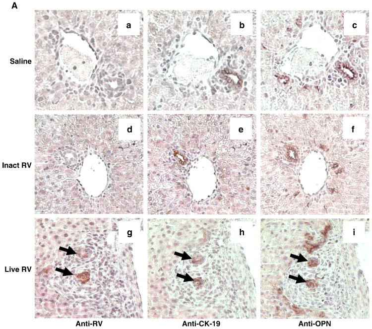
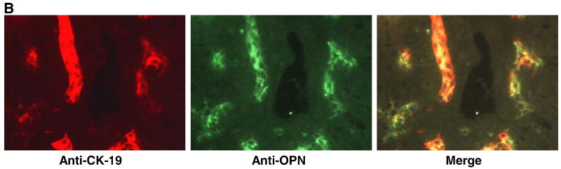
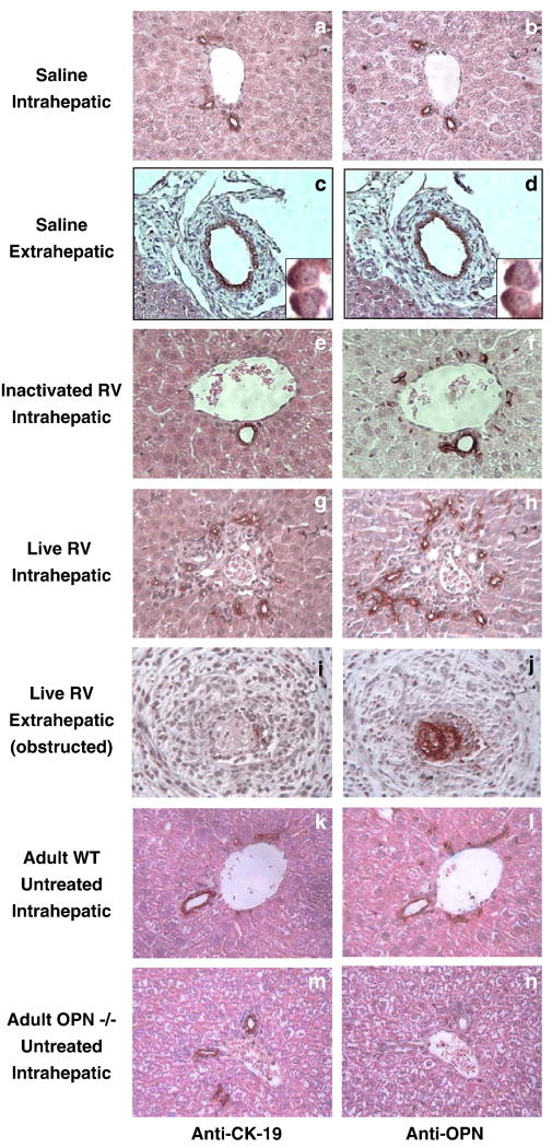
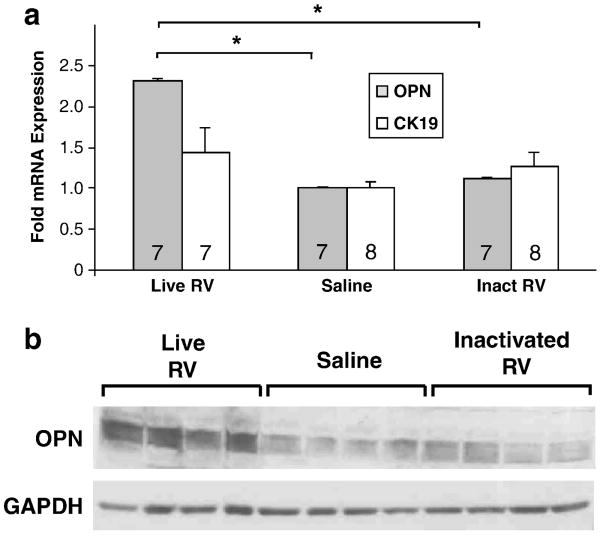
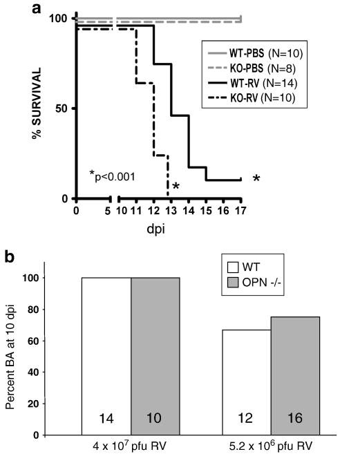
Biliary atresia (BA) is a progressive fibro-inflammatory pediatric liver disease in which osteopontin (OPN), a glycoprotein with inflammatory and fibrogenic activity, may play a pathogenic role. The current studies were conducted in a mouse model of rotavirus-induced BA to test the hypotheses that live but not inactivated rotavirus causes antigenemia, upregulation of hepatic OPN expression, and induction of BA and fibrosis; and that OPN is necessary for development of BA. Prolonged or transient antigenemia developed in mice inoculated with live or inactivated virus, respectively, but only live virus upregulated hepatic OPN and caused BA and fibrosis. OPN was expressed in intra- and extrahepatic bile ducts in healthy mice and in mice with BA. OPN-deficient mice, similar to WT mice, developed BA. Together, these data show that live but not inactivated rotavirus causes upregulation of hepatic OPN expression and BA but that OPN is not necessary for development of BA.


Figures

FIGURE 1
FIGURE 1. Kinetics of Development of BA in Neonatal Mouse Pups
Mice (24-48 hrs after birth) were monitored for the development of BA (acholic stools, bilirubinuria, and, at 14-16 dpi, extrahepatic biliary strictures) at different times after inoculation with purified live triple-layered RRV particles (TLP), inactivated TLP, or saline. Total number of pups evaluated in each group is indicated above each bar. All mice that had acholic stools and bilirubinuria at 15-16 dpi had obvious extrahepatic bile duct and gallbladder abnormalities including strictures and dilatations, reflecting the sensitivity of objective assessment of stool and urine as a tool for detecting BA in this model.
FIGURE 2
FIGURE 2. RV-induced BA in a mouse model shares important clinical characteristics with human BA
(A) Jaundice and stunting of live RV-inoculated mouse with BA. Mouse inoculated with saline (S) weight 11.29g; mouse inoculated with inactivated virus (I) weight 10.93g; and mouse inoculated with live virus (L) weight 5.23g at 16 dpi. Mice (S) and (I) had pigmented stools and normal urine, and mouse (L) had acholic stool,and bilirubinuria. (B) Liver and extrahepatic biliary tree of the same pups under dissecting microscope (∼6×). Live RV-inoculated mouse liver has dark yellow hue, tortuous cystic duct (CD), necrotic foci (arrow) and distended, dark-colored gallbladder (GB) secondary to bile stasis. (C) Live RV-inoculated pups had significantly lower average body mass beginning at 4 dpi (p<0.01) compared with inactivated virus and saline treated pups (*). The average body mass of inactivated RV treated pups was slightly lower than that of saline treated pups, but this did not reach statistical significance until 10 dpi and after (p<0.01) (#).
FIGURE 3
FIGURE 3. Live and Inactivated RV Inoculated Pups Develop Serum RV Antigenemia
RV serum antigenemia at 1, 4, 9-10, and 15-16 dpi. For saline-inoculated pups, N=16, 14, 4, 5, at 4 dpi, 9-10 dpi, and 15-16 dpi, respectively. For inactivated RV-inoculated pups, N=12, 7, 8, and 9 for the same time points. For live RV-inoculated pups, N=4, 7, 7, and 7 for the same time points. The lower limit of antigen detection (0.094) is indicated with a dotted horizontal line. (*) indicates p<0.01 vs saline treatment group. (#) indicates p<0.05 vs saline group. Inactivated RV vs live RV at 1 dpi was NS, and inactivated RV vs saline at 9-10 dpi and at 15-16 dpi was NS.
FIGURE 4
FIGURE 4. Replicating Virus is Present in Bile Ducts of Live RV-Inoculated Pups
Histopathology and RV, NSP4, and CK19 antigen detection in the livers of pups at 4 dpi. Bile ducts of saline (a-c) and inactivated RV (d-f) treated mice do not stain for RV or NSP4. Liver from RV-inoculated pup shows inflammation surrounding foci (arrows) that stain, on consecutive slides, for RV (g), cytokeratin 19 (bile ducts) (h), and the non-structural RV protein NSP4 (i), reflecting the presence of viral replication in bile ducts (20×). Insets show high-magnification images of virus-infected bile ducts, which are also highlighted using arrows (g-i).
FIGURE 5
FIGURE 5. Portal Fibrosis is Evident in Mice Inoculated With Live RV (with BA) at 16 dpi
Healthy inactivated RV-treated mouse and saline control mouse livers have minimal staining limited to areas immediately adjacent to portal structures.The liver of a RV-injected mouse at 16 dpi (20×) stained with picrosirius red shows portal fibrosis (arrows).
FIGURE 6
FIGURE 6. Osteopontin (OPN) is Expressed in RV-Infected and in Uninfected Bile Ducts
(A) Consecutive sections of formalin-fixed mouse liver 4 days post-inoculation with saline (a-c), inactivated RV (d-f), or live RV (g-i) (20×). Areas of RV, CK-19 (bile duct epithelium), and OPN staining are surrounded by dense inflammation in the liver of the live RV-inoculated mouse (g-i); arrows indicate RV-infected bile ducts. Normal bile ducts in saline- and inactivated virus- treated pups also stain for osteopontin in consecutive sections (a-f) in the cytoplasm and on the apical surface, an expression pattern that resembles that of CK-19 (inset c, d). (B) Merged immunofluorescent image (yellow) on frozen liver from a live RV-inoculated mouse with BA at 14 dpi (20×) shows similar staining patterns for CK19 (red) and OPN (green), although expression in some of the CK19-positive cells is relatively weak.
FIGURE 7
FIGURE 7. Osteopontin is Expressed in All Intrahepatic and Extrahepatic Bile Ducts
Consecutive sections of livers 15-16 days post-inoculation with saline (a-d), inactivated RV (e-f), and live RV (g-j) show OPN expression in all intra- and extrahepatic bile ducts, including in an untreated adult mouse (k-l). There is no OPN staining of intrahepatic bile ducts in an OPN-deficient mouse (m-n).
FIGURE 8
FIGURE 8. Osteopontin Expression is Upregulated in Livers of Mice With BA
(A) OPN and CK-19 (bile duct epithelial cell/cholangiocyte marker) mRNA expression in livers of healthy inactivated virus- and saline-inoculated pups compared with livers from live RV-inoculated pups at 14 dpi, all of whom had BA (acholic stools and bilirubinuria) (p<0.05). Number of pups per treatment group indicated inside bars. (*) OPN expression in the livers of live RV-treated mice was significantly higher than in inactivated RV or saline treated mice (p<0.05). (B) OPN protein expression (60-65 kDa, with GAPDH loading control at 37 kDa) in livers of healthy inactivated virus- and saline-inoculated pups compared with livers from live RV-inoculated pups at 14 dpi with BA. Each lane represents the liver of an individual animal.
FIGURE 9
FIGURE 9. OPN Deficiency Does Not Protect Against RV-Induced BA or Improve Survival
(A) Survival curve for WT and OPN-deficient mice inoculated with RV or saline (“PBS”). Survival was significantly longer in RV-inoculated WT mice (N=14), of which two recovered, than in RV-inoculated OPN-/- mice (N=10), of which none recovered (p<0.001). (B) Percent BA in WT and OPN-/- pups inoculated with two different doses of RV. N for each group indicated inside bars. Virus dose on X-axis. A slightly lower percentage of WT pups (67%) developed BA than OPN-/- pups (75%), but this was NS.

Similar articles

Cited by

References

    1. Allen SR, Jafri M, Donnelly B, McNeal M, Witte D, Bezerra J, Ward R, Tiao GM. Effect of rotavirus strain on the murine model of biliary atresia. J Virol. 2007;81(4):1671–9. - PMC - PubMed
    1. Barnes BH, Tucker RM, Wehrmann F, Mack DG, Ueno Y, Mack CL. Cholangiocytes as immune modulators in rotavirus-induced murine biliary atresia. Liver Int. 2008;29(8):1253–61. - PMC - PubMed
    1. Bezerra JA, Tiao G, Ryckman FC, Alonson M, Sabla GE, Shneider B, Sokol RJ, Aronow BJ. Genetic induction of proinflammatory immunity in children with biliary atresia. Lancet. 2002;360:1653–1659. - PubMed
    1. Blutt SE, Fenaux M, Warfield KI, Greenberg HB, Conner ME. Active viremia in rotavirus-infected mice. J Virol. 2006;80(13):6702–6705. - PMC - PubMed
    1. Blutt SE, Matson DO, Crawford SE, Staat MA, Azimi P, Bennett BL, Piedra PA, Conner ME. Rotavirus antigenemia in children is associated with viremia. PLoS Med. 2007;4(4):e121. - PMC - PubMed

Publication types