. 2011 Nov;70(11):2022-8.
doi: 10.1136/ard.2011.153312. Epub 2011 Jul 8.

Cytokine mRNA profiling identifies B cells as a major source of RANKL in rheumatoid arthritis

Affiliations
Free PMC article

Cytokine mRNA profiling identifies B cells as a major source of RANKL in rheumatoid arthritis

Lorraine Yeo et al. Ann Rheum Dis. 2011 Nov.
Free PMC article

Abstract

Objectives: In rheumatoid arthritis (RA), a complex cytokine network drives chronic inflammation and joint destruction. So far, few attempts have been made to identify the cellular sources of individual cytokines systematically. Therefore, the primary objective of this study was systematically to assess the cytokine messenger RNA expression profiles in the five largest cell populations in the synovial fluid and peripheral blood of RA patients. To reflect the in vivo situation as closely as possible, the cells were neither cultured nor stimulated ex vivo.

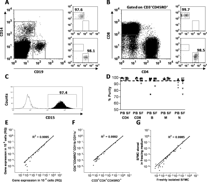
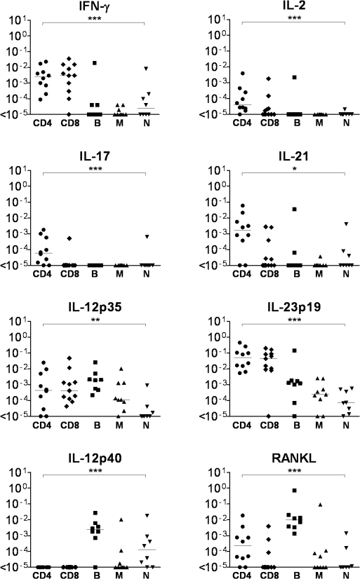
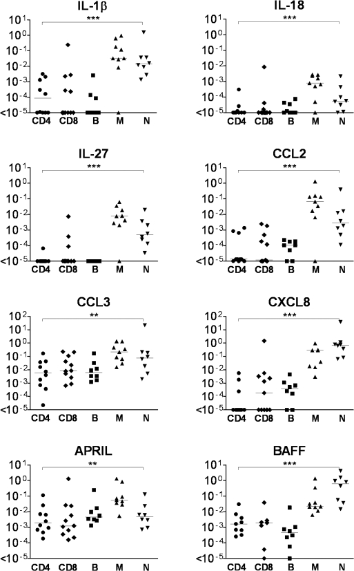
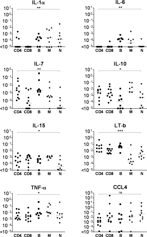
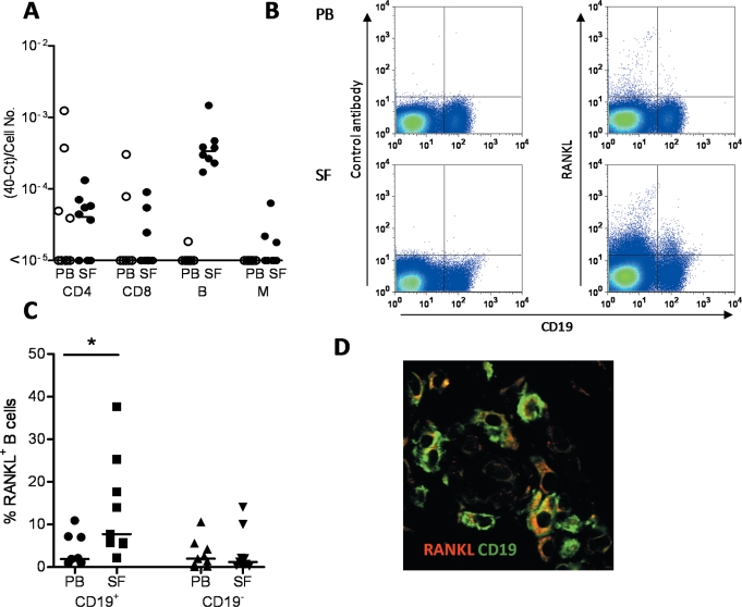
Methods: Inflammatory cells from 12 RA patients were sorted into CD4 and CD8 T cells, B cells, macrophages and neutrophils. mRNA expression for 41 cytokines was determined by real-time PCR using microfluidic cards. Receptor activator nuclear factor kappa B ligand (RANKL) (TNFSF11) expression by B cells was further confirmed by flow cytometry and by immunofluorescence staining of frozen sections of synovial tissue from patients with RA.

Results: The detection of cytokines characteristic for T cells and myeloid cells in the expected populations validated this methodology. Beyond the expected cytokine patterns, novel observations were made. Striking among these was the high expression of mRNA for RANKL in B cells from synovial fluid. This observation was validated at the protein level in synovial tissue and fluid.

Conclusions: RANKL, the key cytokine driving bone destruction by osteoclast activation, is produced by synovial B cells in RA. This observation is of importance for our understanding of the role of B cells in RA and their therapeutic targeting.


Conflict of interest statement

Competing interests None.

Figures

Figure 1
Figure 1
Method validation. (A, B) B cells, monocytes, CD4 T cells and CD8 T cells were sorted based on their expression of CD19, CD14, CD4/CD3/CD45R0 and CD8/CD3/CD45R0, respectively. (C) Neutrophils were isolated with CD15-coated magnetic beads. (D) All samples were re-analysed after the sort and samples with more than 5% contaminating cells were rejected. (E) To establish the cell number needed for the detection of cytokine gene expression, results for 1×10³, 1×10⁴ and 1×10⁵ cells were directly compared in three independent experiments. There was a close correlation between the results yielded by the detection of 43 individual cytokine genes in 1×10⁴ and 1×10⁵ cells, confirming that using a minimum cell number of 104 would be sufficient for the sensitivity of the real-time PCR microfluidic card assay. Comparison of the results from 1×10³ genes and 1×10⁵ cells showed a large degree of variation (data not shown). (F) Comparison of cytokine gene expression for mononuclear cells sorted on the basis of CD3 and CD4 expression compared with T cells sorted on the basis of CD4 expression and lack expression of the myeloid cell markers CD11c and CD11b. Under controlled experimental conditions, keeping the cells below 4°C, T cells sorted using anti-CD3 antibody did not have an altered expression profile compared with T cells sorted using alternative markers, showing that under these conditions there is no induction of cytokine gene expression by the CD3 antibody (data representative of three independent experiments). (G) The cytokine mRNA expression was not altered by storing synovial fluid mononuclear cells in freezing medium at −80°C compared with using freshly isolated cells (data representative of two independent experiments). PB, peripheral blood; SF, synovial fluid; SFMC, synovial fluid mononuclear cell.
Figure 2
Figure 2
Cytokine genes expressed predominantly in synovial fluid lymphoid cells. Shown are the quantitative PCR results for the mRNA expression for a selection of the cytokines predominantly expressed in CD4 T cells, CD8 T cells or in CD19 B cells (B). Statistical significance was assessed by the Kruskal–Wallis test. *p<0.05, **p<0.001, ***p<0.0001.
Figure 3
Figure 3
Cytokine genes expressed predominantly in synovial fluid macrophages or neutrophils. Shown are the quantitative PCR results for the mRNA expression for a selection of the cytokines (N) predominantly expressed in either CD14 monocytes (M) or CD15 neutrophils. Statistical significance was assessed by the Kruskal–Wallis test. **p<0.001, ***p<0.0001.
Figure 4
Figure 4
Cytokines expressed in both synovial fluid myeloid and lymphoid cell populations. Shown are the quantitative PCR results for the mRNA expression for a selection of the cytokines expressed in at least one of the lymphoid cell populations: CD4 T cells, CD8 T cells or in CD19 B cells as well as either CD14 monocytes or CD15 neutrophils. Statistical significance was assessed by the Kruskal–Wallis test. *p<0.05, **p<0.001, ***p<0.0001.
Figure 5
Figure 5
Expression of receptor activator nuclear factor kappa B ligand (RANKL) in B cells infiltrating the rheumatoid synovial tissue and fluid. (A) Quantitative PCR data for detection of RANKL in sorted peripheral blood (PB) and synovial fluid (SF) CD4 and CD8 T cells, CD19 B cells and CD14 monocytes/macrophages. Data normalised to the exact number of cells sorted. (B) Flow cytometric detection of RANKL. Peripheral blood and synovial fluid mononuclear cells were co-labelled with anti-CD19 and either isotype matched control or anti-RANKL antibody. Staining representative of samples from eight patients with rheumatoid arthritis (RA). (C) Percentage of RANKL+ cells among the CD19 B cells of RA patients as detected by flow cytometry. (D) Detection of RANKL+ B cells in synovial tissue. Synovial tissue was stained for CD19 (green), and RANKL (red) and viewed at a final magnification of ×400. Representative sections from five RA patients were investigated.

Comment in

References

    1. Brennan FM, McInnes IB. Evidence that cytokines play a role in rheumatoid arthritis. J Clin Invest 2008;118:3537–45 - PMC - PubMed
    1. Feldmann M, Maini SR. Role of cytokines in rheumatoid arthritis: an education in pathophysiology and therapeutics. Immunol Rev 2008;223:7–19 - PubMed
    1. van den Berg WB. Lessons from animal models of arthritis over the past decade. Arthritis Res Ther 2009;11:250. - PMC - PubMed
    1. Ehrenstein MR, Mauri C. If the treatment works, do we need to know why?: the promise of immunotherapy for experimental medicine. J Exp Med 2007;204:2249–52 - PMC - PubMed
    1. Brennan FM, Chantry D, Jackson A, et al. Inhibitory effect of TNF alpha antibodies on synovial cell interleukin-1 production in rheumatoid arthritis. Lancet 1989;2:244–7 - PubMed

Publication types