. 2011 Aug;22(8):1443-52.
doi: 10.1681/ASN.2010121260. Epub 2011 Jul 8.

IRF4 deficiency abrogates lupus nephritis despite enhancing systemic cytokine production

Affiliations

IRF4 deficiency abrogates lupus nephritis despite enhancing systemic cytokine production

Maciej Lech et al. J Am Soc Nephrol. 2011 Aug.

Abstract

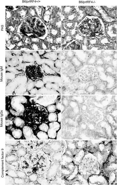
The IFN-regulatory factors IRF1, IRF3, IRF5, and IRF7 modulate processes involved in the pathogenesis of systemic lupus and lupus nephritis, but the contribution of IRF4, which has multiple roles in innate and adaptive immunity, is unknown. To determine a putative pathogenic role of IRF4 in lupus, we crossed Irf4-deficient mice with autoimmune C57BL/6-(Fas)lpr mice. IRF4 deficiency associated with increased activation of antigen-presenting cells in C57BL/6-(Fas)lpr mice, resulting in a massive increase in plasma levels of TNF and IL-12p40, suggesting that IRF4 suppresses cytokine release in these mice. Nevertheless, IRF4 deficiency completely protected these mice from glomerulonephritis and lung disease. The mice were hypogammaglobulinemic and lacked antinuclear and anti-dsDNA autoantibodies, revealing the requirement of IRF4 for the maturation of plasma cells. As a consequence, Irf4-deficient C57BL/6-(Fas)lpr mice neither developed immune complex disease nor glomerular activation of complement. In addition, lack of IRF4 impaired the maturation of Th17 effector T cells and reduced plasma levels of IL-17 and IL-21, which are cytokines known to contribute to autoimmune tissue injury. In summary, IRF4 deficiency enhances systemic inflammation and the activation of antigen-presenting cells but also prevents the maturation of plasma cells and effector T cells. Because these adaptive immune effectors are essential for the evolution of lupus nephritis, we conclude that IRF4 promotes the development of lupus nephritis despite suppressing antigen-presenting cells.


Figures

Figure 1.
Figure 1.
Lack of IRF4 increases the activation of antigen-presenting cells. (A) Serum cytokine levels were determined by ELISA in B6lpr mice (black bars) and B6lpr/Irf4−/− mice (white bars) at 6 months of age. (B) Spleen monocytes were stimulated with 1 μg/ml LPS and the cytokine levels were determined by ELISA. (C-E) Spleen cells were quantified by flow cytometry using surface activation markers as indicated. Data represent mean ± SEM from 12 to 14 mice in each group. *P < 0.05, **P < 0.01 versus B6lpr mice.
Figure 2.
Figure 2.
Lack of IRF4 abrogates renal pathology in 6-month-old B6lpr mice. Renal sections from 6-month-old B6lpr and B6lpr/Irf4−/− mice were stained with periodic acid–Schiff (PAS), anti-mIgM, anti-mIgG, or anti-complement factor C9 as indicated. Note that B6lpr but not B6lpr/Irf4−/− mice develop diffuse proliferative glomerulonephritis with IgM, IgG, and C9 deposits. Original magnification, ×400.
Figure 3.
Figure 3.
Lack of IRF4 abrogates lupus nephritis in 6-month-old B6lpr mice. The indices of lupus nephritis disease activity and chronicity were assessed by semiquantitative morphometry on PAS-stained renal sections from 6-month-old mice of both mouse strains as described in Concise Methods. Plasma creatinine levels were determined at the same age. Data represent mean ± SEM from at least 12 mice per group.
Figure 4.
Figure 4.
Lack of IRF4 reduces renal chemokine mRNA expression in 6-month-old B6lpr mice. (A) Renal mRNA was isolated from 6-month-old B6lpr mice (black bars) and B6lpr/Irf4−/− mice (white bars) and quantified by real-time reverse-transcriptase PCR. Data are expressed as mean of the ratio versus the respective 18S rRNA level ± SEM. (B) Mac2-positive macrophages were quantified in 20 glomeruli per section in both mouse strains at 6 months of age. Data represent mean ± SEM from at least 12 mice per group. *P < 0.05 versus B6lprmice.
Figure 5.
Figure 5.
Lack of IRF4 abrogates lung disease in 6-month-old B6lpr mice. (A) Lung sections from 6-month-old B6lpr and B6lpr/Irf4−/− mice were stained with PAS. Note that B6lpr but not B6lpr/Irf4−/− mice develop peribronchial and perivascular immune cell infitrates. Original magnification, ×100. (B) Mac2-positive glomerular cells were quantified in 20 glomeruli per section. Data are mean cell counts per glomerulus ± SEM of 9 to 12 mice in each group. ***P < 0.001 versus B6lpr mice.
Figure 6.
Figure 6.
IRF4 deficient B6lpr mice lack hypergammaglobuminemia and autoantibody production. (A) B6lpr/Irf4−/− (○) and B6lpr wild-type mice (●) were bled at the end of the study to determine serum levels of IgG, IgM, anti-Smith, and anti-dsDNA autoantibodies by ELISA. (B) ANAs were detected by staining of Hep2 cells using plasma dilutions of 1:40 as described in Concise Methods. Note the homogenous nuclear staining pattern using plasma from B6lprmice that was not detectable with plasma from B6lpr/Irf4−/− mice.
Figure 7.
Figure 7.
IRF4 is required for plasma cells' maturation. Flow cytometry was used to determine the total number of distinct B and plasma cell subsets (A) in spleens of 6-month-old B6lpr mice (black bars) and B6lpr/Irf4−/− mice (white bars). The histogram presents mean ± SEM of 12 to 14 mice in each group. *P < 0.05, ***P < 0.001 versus B6lpr mice. (B) IgM immunostaining of spleens identifies mature B cell distribution in mice of both genotypes. Original magnification, ×100.
Figure 8.
Figure 8.
Lack of IRF4 impairs the maturation of Th17 cells. Flow cytometry was used to determine (A) T cell subsets and IFNγ- and (B) IL-17-producing CD4 T cells in spleens of 6-month-old B6lpr mice (black bars) and B6lpr/Irf4−/− mice (white bars). (C) Plasma IL-17 and IL-21 levels were determined by ELISA at 6 months of age. The histogram presents mean ± SEM of 12 to 14 mice in each group. *P < 0.05, **P < 0.01 versus B6lpr mice.

Similar articles

Cited by

References

    1. Kotzin BL: Systemic lupus erythematosus. Cell 85: 303–306, 1996 - PubMed
    1. Goodnow CC: Multistep pathogenesis of autoimmune disease. Cell 130: 25–35, 2007 - PubMed
    1. Gregersen PK, Olsson LM: Recent advances in the genetics of autoimmune disease. Annu Rev Immunol 27: 363–391, 2009 - PMC - PubMed
    1. Kanta H, Mohan C: Three checkpoints in lupus development: Central tolerance in adaptive immunity, peripheral amplification by innate immunity and end-organ inflammation. Genes Immun 10: 390–396, 2009 - PubMed
    1. Theofilopoulos AN, Baccala R, Beutler B, Kono DH: Type I interferons (alpha/beta) in immunity and autoimmunity. Annu Rev Immunol 23: 307–336, 2005 - PubMed

Publication types

Substances