. 2011 Oct;39(19):8572-85.
doi: 10.1093/nar/gkr560. Epub 2011 Jul 8.

Structural basis for the biological relevance of the invariant apical stem in IRES-mediated translation

Affiliations

Structural basis for the biological relevance of the invariant apical stem in IRES-mediated translation

Noemí Fernández et al. Nucleic Acids Res. 2011 Oct.

Abstract

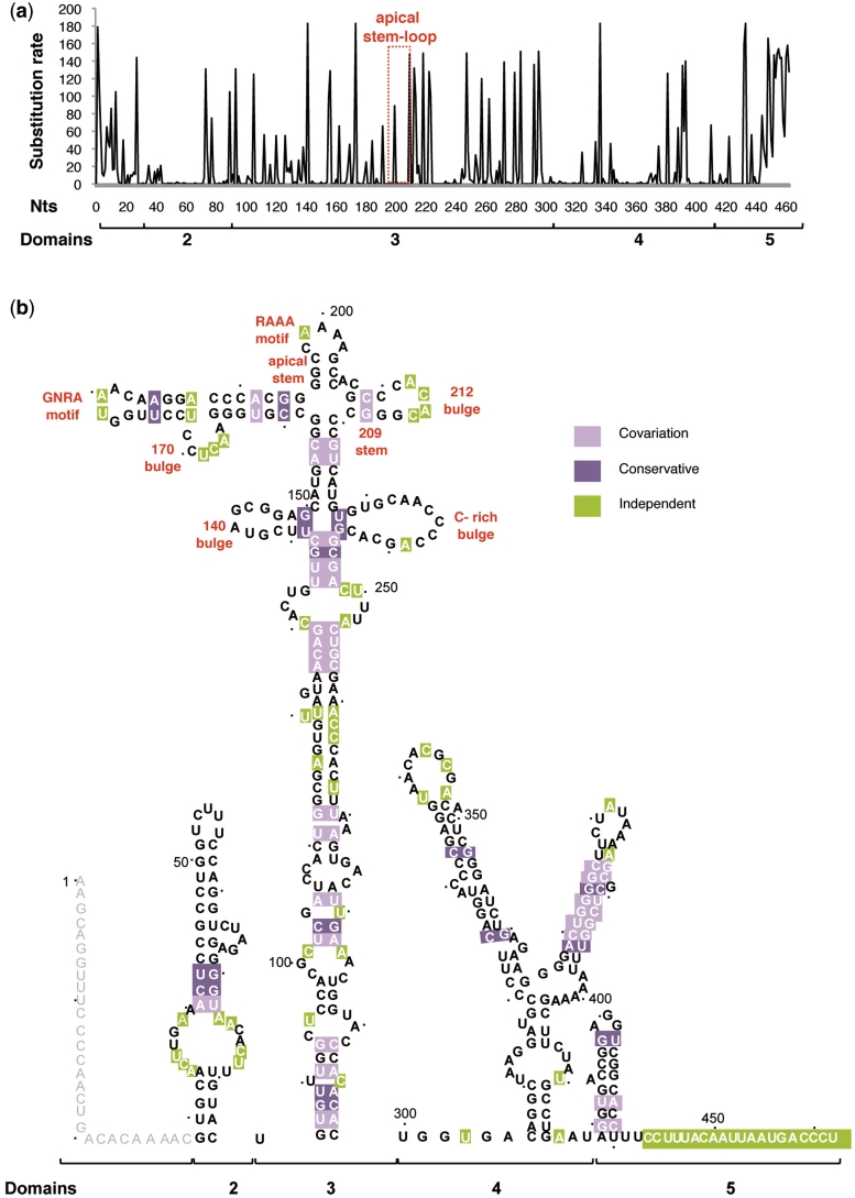
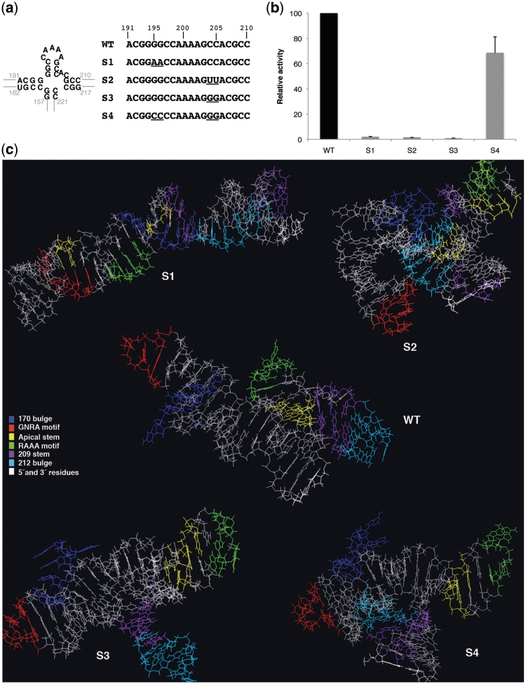
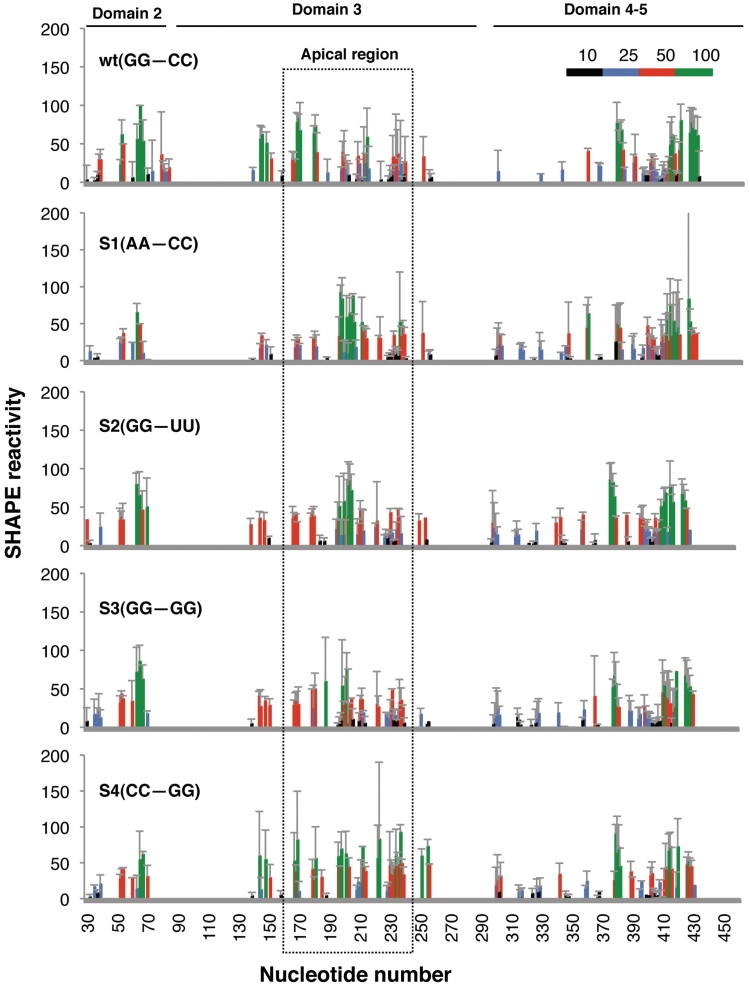
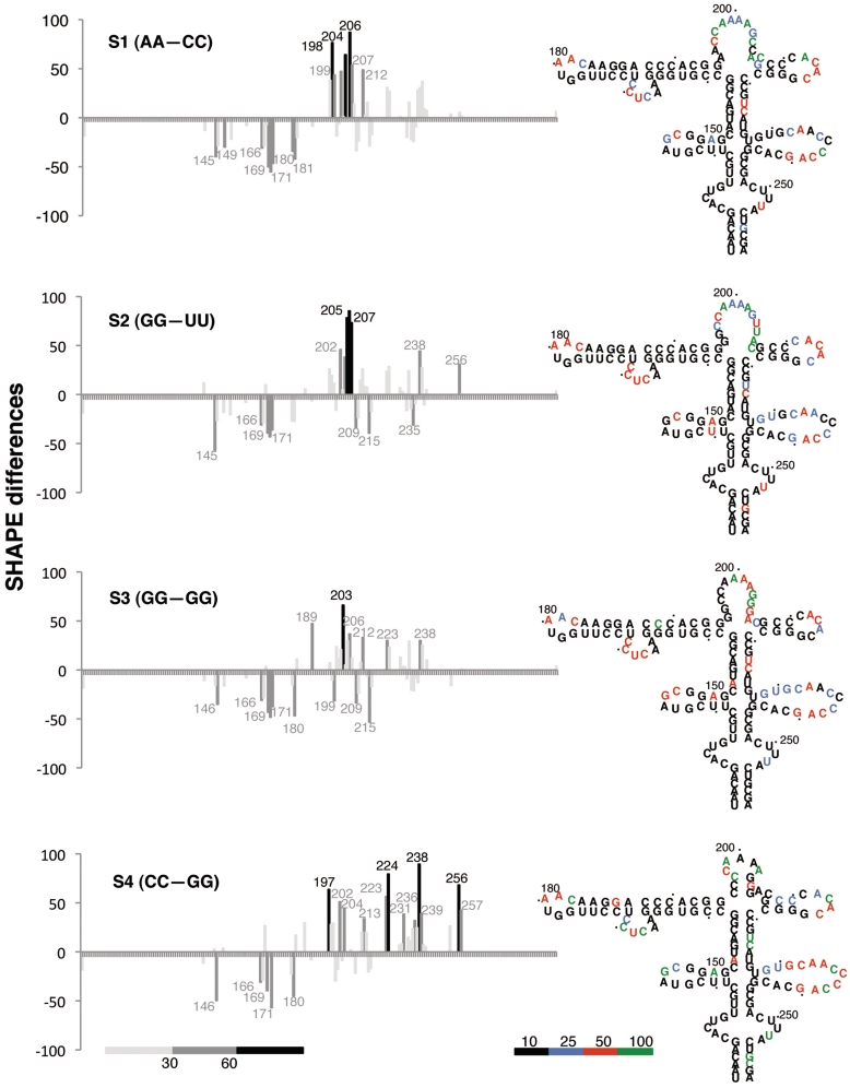
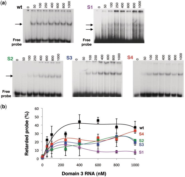
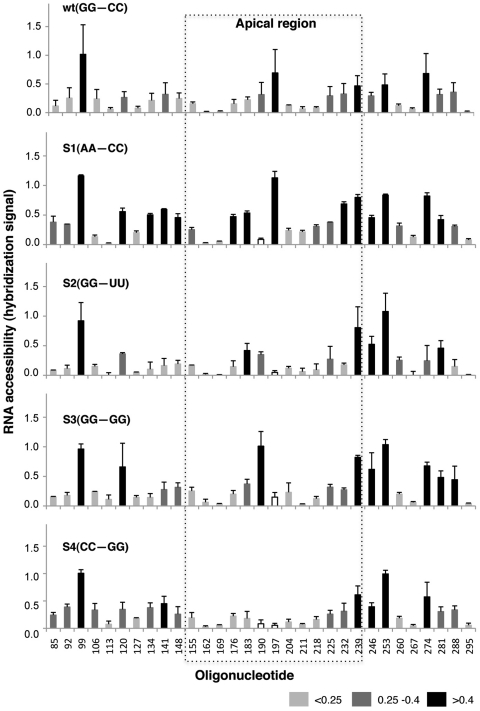
RNA structure plays a fundamental role in internal initiation of translation. Picornavirus internal ribosome entry site (IRES) are long, efficient cis-acting elements that recruit the ribosome to internal mRNA sites. However, little is known about long-range constraints determining the IRES RNA structure. Here, we sought to investigate the functional and structural relevance of the invariant apical stem of a picornavirus IRES. Mutation of this apical stem revealed better performance of G:C compared with C:G base pairs, demonstrating that the secondary structure solely is not sufficient for IRES function. In turn, mutations designed to disrupt the stem abolished IRES activity. Lack of tolerance to accept genetic variability in the apical stem was supported by the presence of coupled covariations within the adjacent stem-loops. SHAPE structural analysis, gel mobility-shift and microarrays-based RNA accessibility revealed that the apical stem contributes to maintain IRES RNA structure through the generation of distant interactions between two adjacent stem-loops. Our results demonstrate that a highly interactive structure constrained by distant interactions involving invariant G:C base pairs plays a key role in maintaining the RNA conformation necessary for IRES-mediated translation.


Figures

Figure 1.
Figure 1.
Sequence variability of the IRES element in FMDV RNA. (a) The total number of changes found in 183 aligned IRES sequences is plotted against the nucleotide position. Location of the residues conforming the apical stem–loop, including the variable R residue of the RAAA motif, is indicated by a rectangle. (b) Sequence heterogeneity maintains the secondary structure of the FMDV IRES. Invariant nucleotides in the IRES secondary structure are marked in bold. Covariant and conservative nucleotide changes are depicted by light and dark violet squares, respectively. Independent substitutions are marked by green squares. Nucleotide positions are denoted by dots 10 nt apart. The most relevant motifs referred to in the text are indicated. Distribution of the IRES in domains (2–5) is indicated below each panel.
Figure 2.
Figure 2.
Mutational analysis of the invariant apical stem. (a) Nucleotide substitutions present in S1–S4 mutants are represented below the wild-type C-S8 FMDV IRES sequence (nucleotides 191–210). A diagram of the wild-type secondary structure of this region is shown (left). (b) Relative IRES activity, determined as the ratio of luciferase to CAT in BHK-21 cells transfected with plasmids of the form CAT-IRES-luciferase made relative to the activity obtained with the wt IRES. Values correspond to the mean of three independent assays. Errors bars, SD. (c) PDB RNA structure prediction by Mc-fold pipeline of the apical region of domain 3 (nucleotides 155–223) bearing wt and S1–S4 mutant sequences. Location of the loops and stems referred to in the text are depicted by the color code listed in the legend.
Figure 3.
Figure 3.
SHAPE reactivity of apical stem IRES mutants. Values of SHAPE reactivity at each individual nucleotide position correspond to the mean reactivity (±SD) of three independent assays. RNAs, treated with NMIA or untreated, were subjected to primer extension analysis conducted with 5′-end labeled primers. The intensity of each band was normalized to the full-length cDNA product detected in the corresponding gel lane after subtraction of the corresponding background RT-stop signal in the untreated RNA. Nucleotide positions are indicated on the x-axis. The SHAPE reactivity is depicted using color-coded bars. Position of the apical region is indicated by a broken line rectangle.
Figure 4.
Figure 4.
Impact of the apical stem sequence composition on SHAPE reactivity differences. SHAPE difference plots of each mutant domain 3 relative to the wt RNA. Nucleotides with absolute changes in SHAPE reactivity >60% are depicted in black, while those between 30 and 60% are marked in dark grey. The secondary structure model of each IRES mutant, with nucleotides colored as in Figure 3 to reflect their mean SHAPE reactivity, is shown on the right. The reference mean SHAPE reactivity of the wt RNA is shown in Supplementary Figure S1.
Figure 5.
Figure 5.
Mobility-shift binding assays of domain 3 RNAs with the GNRA hairpin. (a) Representative examples of gel-shift assays for each construct, wt and S1 to S4 mutant RNAs. Gel-shift assays were carried out using 32P-labeled GNRA hairpin (corresponding to nucleotides 160–196 of domain 3) (50 nM) as probe and the wild-type or mutant unlabeled domain 3 RNAs (0–1000 nM). The position of the retarded complex is depicted by an arrow. Note that a longer exposure is shown for S1 RNA to allow the detection of two weak retarded complexes. (b) The percentage of retarded probe calculated from duplicate assays was plotted against the unlabeled RNA concentration. Error bars, SD.
Figure 6.
Figure 6.
Hybridization of IRES transcripts to antisense oligonucleotides printed on microarrays. Hybridization signal of the fluorescent-labeled domain 3 transcripts plotted against each oligonucleotide (mean ± SD) averaged from three independent assays. The array contains 14-nt long oligonucleotides (termed 1–449, by the position on the IRES sequence complementary to the 3′-end of the primer), overlapping each 7 nt within domain 3 (34). Changes in accessibility of mutant IRES to oligonucleotides 190 (S1, S4) and 197 (S2, S3, S4), depicted with white bars, are likely due to mismatches in the sequence of these RNAs with the sequence printed in the microarray. Position of the apical region is indicated by a broken line rectangle. Differences in accessibility are depicted in black (>0.4), grey (range 0.4–0.25) and light grey (<0.25).

Similar articles

Cited by

References

    1. Sonenberg N, Hinnebusch AG. Regulation of translation initiation in eukaryotes: mechanisms and biological targets. Cell. 2009;136:731–745. - PMC - PubMed
    1. Martinez-Salas E. The impact of RNA structure on picornavirus IRES activity. Trends Microbiol. 2008;16:230–237. - PMC - PubMed
    1. Spriggs KA, Bushell M, Willis AE. Translational regulation of gene expression during conditions of cell stress. Mol. Cell. 2010;40:228–237. - PubMed
    1. Costantino DA, Pfingsten JS, Rambo RP, Kieft JS. tRNA-mRNA mimicry drives translation initiation from a viral IRES. Nat. Struct. Mol. Biol. 2008;15:57–64. - PMC - PubMed
    1. Vallejos M, Deforges J, Plank TD, Letelier A, Ramdohr P, Abraham CG, Valiente-Echeverria F, Kieft JS, Sargueil B, Lopez-Lastra M. Activity of the human immunodeficiency virus type 1 cell cycle-dependent internal ribosomal entry site is modulated by IRES trans-acting factors. Nucleic Acids Res. 2011 April 10; epub ahead of print; doi:10.1093/nar/gkr189. - PMC - PubMed

Publication types