. 2011 Oct;39(19):8329-41.
doi: 10.1093/nar/gkr529. Epub 2011 Jul 8.

Histone variant H3.3 stimulates HSP70 transcription through cooperation with HP1γ

Affiliations

Histone variant H3.3 stimulates HSP70 transcription through cooperation with HP1γ

Hyunjung Kim et al. Nucleic Acids Res. 2011 Oct.

Abstract

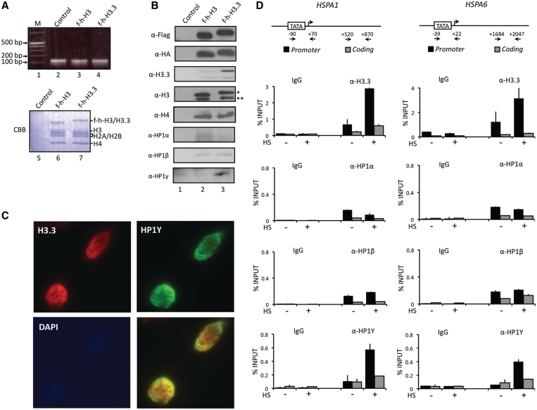
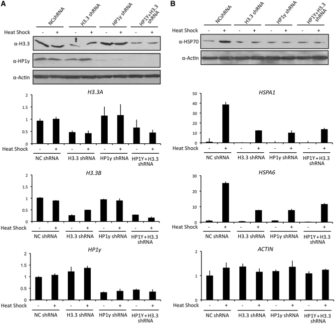
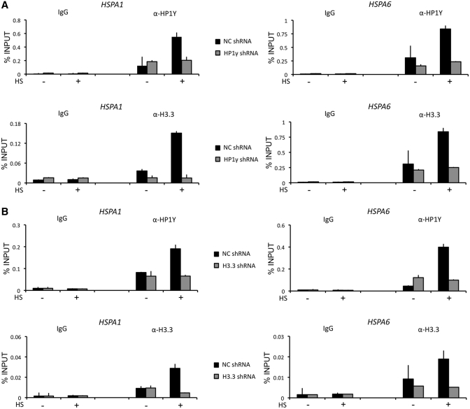
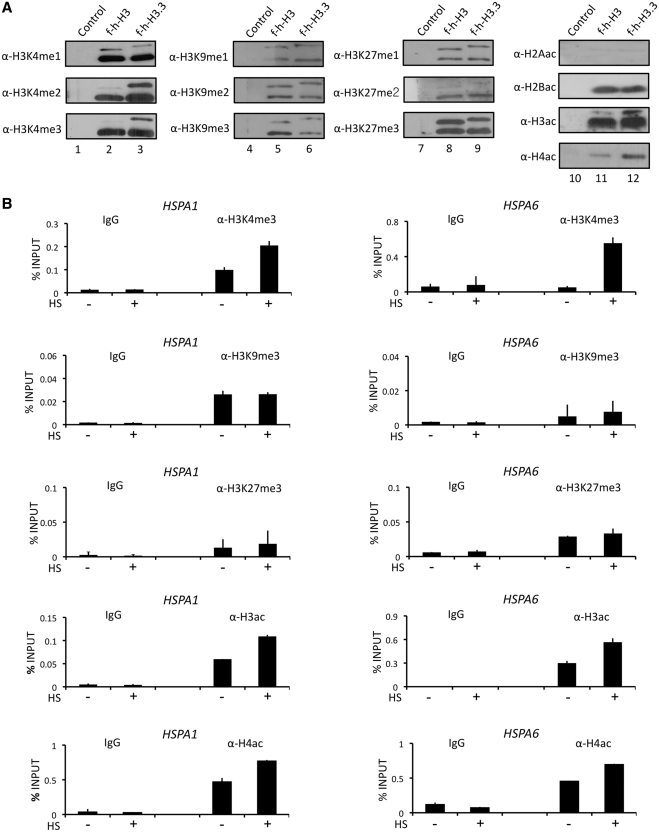
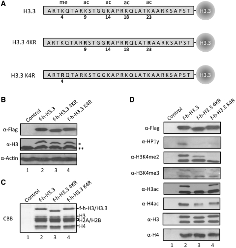
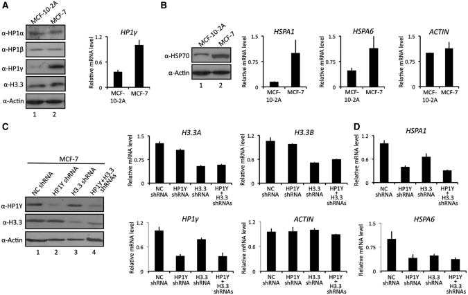
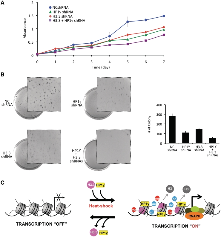
Histone variant H3.3 and heterochromatin protein 1γ (HP1γ) are two functional components of chromatin with role in gene transcription. However, the regulations of their dynamics during transcriptional activation and the molecular mechanisms underlying their actions remain poorly understood. Here, we provide evidence that heat shock-induced transcription of the human HSP70 gene is regulated via the coordinated and interdependent action of H3.3 and HP1γ. H3.3 and HP1γ are rapidly co-enriched at the human HSP70 promoters upon heat shock in a manner that closely parallels the initiation of transcription. Knockdown of H3.3 prevents the stable recruitment of HP1γ, inhibits active histone modifications, and attenuates HSP70 promoter activity. Likewise, knockdown of HP1γ leads to the decreased levels of H3.3 in the promoter regions and the repression of HSP70 genes. HP1γ selectively recognizes particular modification states of H3.3 in the nucleosome for its action. Moreover, HP1γ is overexpressed in three representative cancer cell lines, and its knockdown leads to reduction in HSP70 gene transcription and inhibition of cancer cell proliferation. We conclude that the physical and functional interactions between H3.3 and HP1γ make a unique contribution to acute HSP70 transcription and cancer development related to the misregulation of this transcription event.


Figures

Figure 1.
Figure 1.
Selective interaction of HP1γ with H3.3 nucleosomes. (A) HeLa cells were transfected with control (lane 2), histone H3 (lane 3) and H3.3 (lane 4) expression vectors for 48 h, and mononucleosomes were prepared as summarized in Supplementary Figure S1A. Total nucleosomal DNA was subjected to 2% agarose gel electrophoresis and visualized with ethidium bromide (lanes 2–4). Mononucleosomes containing H3 or H3.3 were purified by sequential immunoprecipitations using anti-Flag and anti-HA antibodies, and histone compositions of the purified nucleosomes were analyzed by Coomassie staining following 15% SDSPAGE (lanes 6 and 7). Lane 1, 0.1–12 kb DNA ladder; lane 5, control mock-purified material. (B) Proteins co-purified with H3 and H3.3 nucleosomes were separated by 15% SDS–PAGE, and the presence of indicated proteins were analyzed by Western blotting. Lane 1, mock-purified material; lane 2, H3 mononucleosomes; lane 3, H3.3 mononucleosomes. Asterisk indicates ectopic f-h-H3 or f-h-H3.3 proteins and double asterisks indicate endogenous H3 or H3.3 proteins. (C) HeLa cells were immunostained for HP1γ (green channel), H3.3 (red channel) and DAPI (blue channel). Image overlays show co-localization between HP1γ and H3.3. (D) HeLa cells were mock-treated (−) or heat-treated (+) for 30 min, and quantitative chromatin immunoprecipitation (ChIP) assays of promoter (filled square) and coding (shaded square) regions of HSPA1 and HSPA6 genes were performed using antibodies specifically recognizing H3.3, HP1α, HP1β and HP1γ. IgG antibody was used as a negative control. Input DNA and immunoprecipitated DNA were analyzed by qPCR analyses using primer sets depicted in the top panel. The results are shown as percentage of input. The error bar indicates the means ± SD.
Figure 2.
Figure 2.
Requirements for H3.3 and HP1γ in HSP70 transcription. (A) HeLa cells were stably transfected with shRNAs targeting to non-specific control, H3.3 or HP1γ. The efficiency and selectivity of knockdown were determined under non-heat shock and heat shock conditions by western blotting (upper panel) and qRT–PCR (lower panel). All transcription levels were normalized to that of GAPDH. Note that H3.3 protein is encoded by two different genes, H3.3 A and H3.3B. β-Actin was used as an internal control for equal loading in all western blot analyses. (B) Whole-cell extracts were prepared for analyses of HSP70 expression in these cells by western blotting analysis. Total mRNA was purified and subjected to qRT–PCR to examine transcription levels of HSPA1 and HSPA6 genes under non-heat shock and heat shock conditions. Average and SD of three independent experiments are shown.
Figure 3.
Figure 3.
Interdependent localization of H3.3 and HP1γ at HSP70 promoters. (A) Mock-depleted (filled square) and H3.3-depleted (shaded square) cells were heat-treated as in Figure 1D, and ChIP assays of HSP70 promoter regions were performed using antibodies against H3.3 and HP1γ. (B) ChIP assays were essentially identical to Figure 3A, but in H3.3-depleted cells.
Figure 4.
Figure 4.
Enrichment of active modifications in H3.3 nucleosomes. (A) Mononucleosomes containing ectopic H3 or H3.3 were prepared as in Figures 1A and S1A, and analyzed by western blotting using antibodies that recognize H3–K4 methylation (α-H3K4me1/me2/me3) H3-K9 methylation (α-H3K9me1/me2/me3), H3–K27 methylation (α-H3K27me1/me2/me3), H2A acetylation (α-H2Aac), H2B acetylation (α-H2Bac), H3 acetylation (α-H3ac) and H4 acetylation (α-H4ac). Lanes 1, 4, 7 and 10, mock-purified materials; lanes 2, 5, 8 and 11, H3 mononucleosomes; lanes 3, 6, 9 and 12, H3.3 mononucleosomes. (B) Cells were mock-treated (−) or heat-treated (+) for 30 min, and modification status of promoter nucleosomes was determined. ChIP assays were essentially as described in Figure 3D.
Figure 5.
Figure 5.
Preferential binding of HP1γ to H3.3 containing mononucleosomes with active modifications. (A) Schematic diagrams of wild type (H3.3), acetylation site-mutated (H3.3 4KR) and K4-mutated (H3.3 K4R) H3.3. Four acetylatable lysine residues (K9, K14, K18 and K23) and one methylatable lysine residue (K4) are mutated to arginine. (B) HeLa cells were transfected with control (lane 1), wild type (lane 2) and mutant H3.3 (lane 3 and 4) expression plasmids for 48 h, and the expression levels of ectopic H3.3 were confirmed by western blotting. (C) Mononucleosomes containing wild type and mutant H3.3 were immunoprecipitated from total mononucleosomes, essentially following the procedure employed in Figure 1. Histone compositions of the purified nucleosomes were analyzed by 15% SDS–PAGE followed by Coomassie staining. (D) H3.3 mononucleosomes were purified as in Supplementary Figure S1A, and analyzed by western blotting using antibodies that recognize H3 acetylation (α-H3ac), H4 acetylation (α-H4ac), H3K4 dimethylation (α-H3K4me2), H3K4 trimethylation (α-H3K4me3), H3 (α-H3), H4 (α-H4) and HP1γ (α-HP1γ). Asterisk indicates ectopic f-h-H3 or f-h-H3.3 proteins and double asterisks indicate endogenous H3 or H3.3 proteins.
Figure 6.
Figure 6.
Affects of H3.3/HP1γ knockdown on HSP70 transcription. (A) MCF-7 breast cancer cells and untransformed MCF-10-2 A breast epithelial cells were subjected to western blot analysis using antibodies specific for HP1α, HP1β, HP1γ and H3.3 (left panel). β-Actin served as a control for equal protein loading. qRT–PCR was performed as in Figure 2A (right panel). (B) HSP70 expression was assessed by western blot with anti-HSP70 antibody (left panel). Transcription of HSPA1 and HSPA6 genes in MCF-10-2 A and MCF-7 cells was quantified by qRT–PCR and corrected for expression of the control gene (GAPDH) (right panel). qRT–PCR was also performed to measure β-Actin mRNA expression (ACTIN). (C) MCF-7 cells were transfected with shRNAs targeting H3.3 and/or HP1γ, and individual and simultaneous depletions of H3.3 and HP1γ were confirmed by western blot analysis (left panel) and qRT–PCR (center and right panels). (D) Transcription levels of HSPA1 and HSPA6 genes of H3.3/HP1γ-depleted MCF-7 cells were analyzed as in Figure 6B.
Figure 7.
Figure 7.
H3.3/HP1γ knockdown-induced alterations in cancer cell growth. (A) MCF-7 cells were depleted of H3.3 and/or HP1γ as in Figure 6C and cell proliferation was measured by MTT assay. Results represent the mean ± SD of three experiments performed in triplicate. (B) H3.3/HP1γ-depleted MCF-7 cells were subjected to soft agar colony formation assay. The graph illustrates the total number of colonies present on the plate after three weeks of culture. Error bars on the graph indicate the standard deviation from triplicate experiments. (C) Models showing the combinatorial role of H3.3 and HP1γ in heat shock-induced HSP70 transcription. Upon heat shock, H3.3 is incorporated at HSP70 promoters and contributes to rapid changes in active histone modifications to stimulate the stable localization of HP1γ at the promoter nucleosomes. The co-enrichment of H3.3 and HP1γ converts HSP70 gene from a repressor state to an active state, leading to a great increase in transcription of HSP70 genes. Lack of H3.3 nucleosomes disrupts the recruitment of HP1γ, which drives the equilibrium further toward H3.3 dissociation, resulting in blockage of transcription initiation. See the ‘Discussion’ section for more details.

Similar articles

Cited by

References

    1. Kouzarides T. Chromatin modifications and their function. Cell. 2007;128:693–705. - PubMed
    1. Henikoff S. Nucleosome destabilization in the epigenetic regulation of gene expression. Nat. Rev. Genet. 2008;9:15–26. - PubMed
    1. Wells D, Hoffman D, Kedes L. Unusual structure, evolutionary conservation of non-coding sequences and numerous pseudogenes characterize the human H3.3 histone multigene family. Nucleic Acids Res. 1987;15:2871–2889. - PMC - PubMed
    1. Albig W, Bramlage B, Gruber K, Klobeck HG, Kunz J, Doenecke D. The human replacement histone H3.3B gene (H3F3B) Genomics. 1995;30:264–272. - PubMed
    1. Tagami H, Ray-Gallet D, Almouzni G, Nakatani Y. Histone H3.1 and H3.3 complexes mediate nucleosome assembly pathways dependent or independent of DNA synthesis. Cell. 2004;116:51–61. - PubMed

Publication types

MeSH terms