Review
. 2011 Jul;91(3):1071-121.
doi: 10.1152/physrev.00038.2010.

Normalization of the vasculature for treatment of cancer and other diseases

Affiliations
Review

Normalization of the vasculature for treatment of cancer and other diseases

Shom Goel et al. Physiol Rev. 2011 Jul.

Erratum in

  • Physiol Rev. 2014 Apr;94(2):707

Abstract

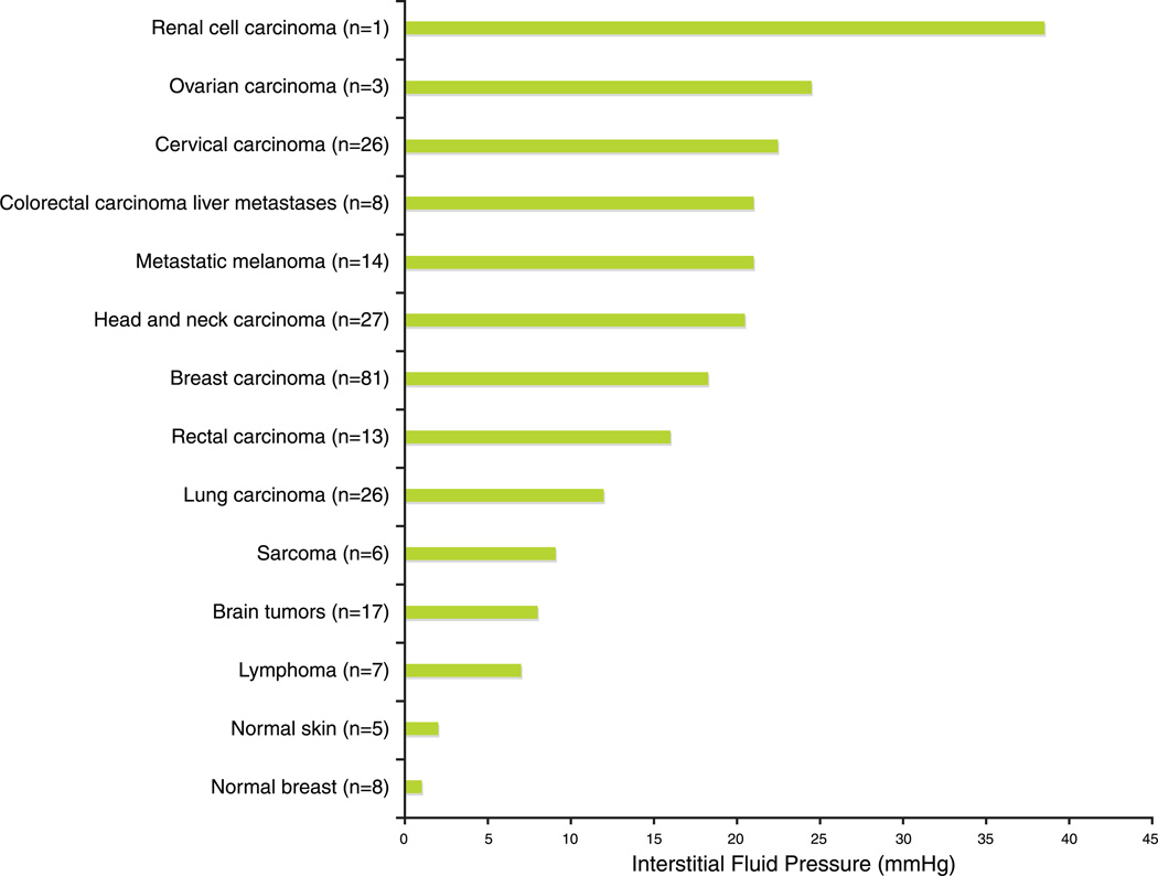
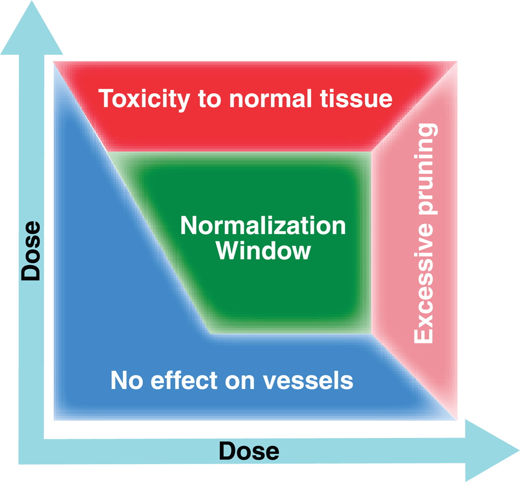
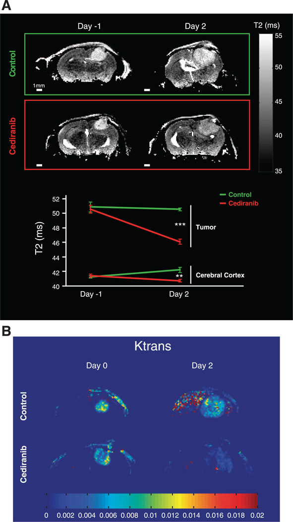
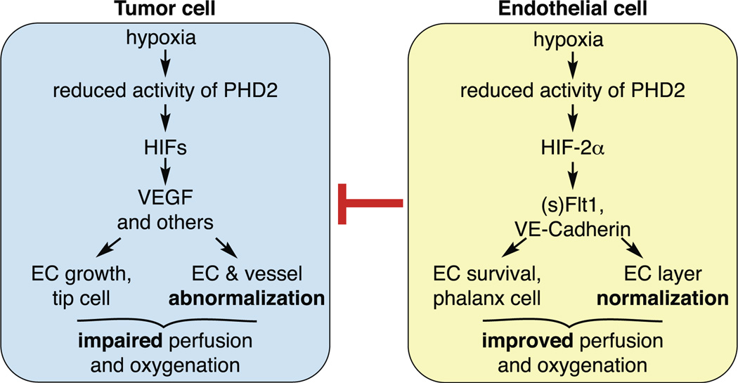
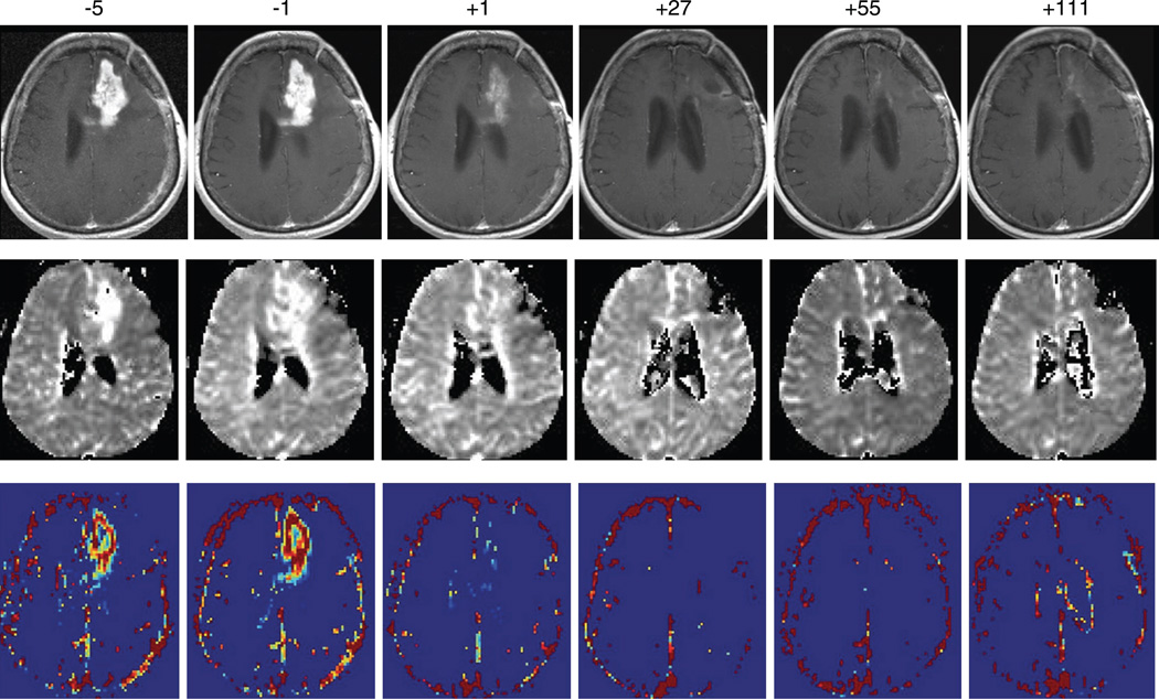
New vessel formation (angiogenesis) is an essential physiological process for embryologic development, normal growth, and tissue repair. Angiogenesis is tightly regulated at the molecular level. Dysregulation of angiogenesis occurs in various pathologies and is one of the hallmarks of cancer. The imbalance of pro- and anti-angiogenic signaling within tumors creates an abnormal vascular network that is characterized by dilated, tortuous, and hyperpermeable vessels. The physiological consequences of these vascular abnormalities include temporal and spatial heterogeneity in tumor blood flow and oxygenation and increased tumor interstitial fluid pressure. These abnormalities and the resultant microenvironment fuel tumor progression, and also lead to a reduction in the efficacy of chemotherapy, radiotherapy, and immunotherapy. With the discovery of vascular endothelial growth factor (VEGF) as a major driver of tumor angiogenesis, efforts have focused on novel therapeutics aimed at inhibiting VEGF activity, with the goal of regressing tumors by starvation. Unfortunately, clinical trials of anti-VEGF monotherapy in patients with solid tumors have been largely negative. Intriguingly, the combination of anti-VEGF therapy with conventional chemotherapy has improved survival in cancer patients compared with chemotherapy alone. These seemingly paradoxical results could be explained by a "normalization" of the tumor vasculature by anti-VEGF therapy. Preclinical studies have shown that anti-VEGF therapy changes tumor vasculature towards a more "mature" or "normal" phenotype. This "vascular normalization" is characterized by attenuation of hyperpermeability, increased vascular pericyte coverage, a more normal basement membrane, and a resultant reduction in tumor hypoxia and interstitial fluid pressure. These in turn can lead to an improvement in the metabolic profile of the tumor microenvironment, the delivery and efficacy of exogenously administered therapeutics, the efficacy of radiotherapy and of effector immune cells, and a reduction in number of metastatic cells shed by tumors into circulation in mice. These findings are consistent with data from clinical trials of anti-VEGF agents in patients with various solid tumors. More recently, genetic and pharmacological approaches have begun to unravel some other key regulators of vascular normalization such as proteins that regulate tissue oxygen sensing (PHD2) and vessel maturation (PDGFRβ, RGS5, Ang1/2, TGF-β). Here, we review the pathophysiology of tumor angiogenesis, the molecular underpinnings and functional consequences of vascular normalization, and the implications for treatment of cancer and nonmalignant diseases.


Figures

FIGURE 1
FIGURE 1
Top: the microvasculature of solid tumors demonstrates a number of structural abnormalities when compared with that of healthy tissues. As shown in A, endothelial cells demonstrate aberrations in shape and are often separated by wide and irregular interendothelial junctions. In addition, there are fewer pericytes, which are often loosely attached to endothelial cells and lie within a basement membrane that is either abnormally thin or thick. The widened endothelial junctions, coupled with the more tenuous vascular investment by pericytes, promote vascular hyperpermeability and facilitate the intravasation of tumor cells into the circulation, such that they can disseminate to form distant metastases. The accompanying functional derangements in tumor microvessels create a hostile microenvironment that fuels tumor growth, metastasis, and resistance to therapies. B: preclinical and clinical data support the notion that anti-angiogenic therapies can “normalize” the tumor vasculature, restoring the structural and functional aberrations of vessels towards a more normal state. Bottom: abnormalities in perivascular cell (PVC) morphology in solid tumors. A: PVCs (stained for α SMA) in normal pancreatic vessels provide a circumferential and tight investment of arterioles and venules in the normal pancreas, with a more dense arrangement around the arteriole. B: double staining for PVCs and endothelial cells (ECs, stained for CD31) in a smaller arteriole and venule in the pancreas shows a regular arrangement of PVCs around the arteriole, with more irregularly arranged and shaped PVCs around the venule. C: PVCs (stained for desmin) on a normal pancreatic capillary are arranged longitudinally along the vessel axis. D and E: PVCs in the MCa-IV mammary carcinoma show morphological abnormalities (stained for αSMA in D and desmin in E) including an irregular arrangement and cytoplasmic projections in multiple directions. F: PVCs in the Lewis Lung Carcinoma (LLC) show loose associations with ECs. G and H: PVCs in the MCa-IV carcinoma and LLC showing occasional PVC-PVC contact and overlap. [Scale bar in H applies to all panels. Bar lengths: 35 µm (A, F–H); 30 µm (B); 15 µm (C); 80 µm (D and E).] [Top panel from Jain RK. N Engl J Med 360: 2669–2671, 2009, with permission, copyright MMS; bottom panel from Morikawa et al. (206), with permission.]
FIGURE 2
FIGURE 2
Anatomical abnormalities in the vascular network of solid tumors, as demonstrated in colorectal carcinoma. A: vascular anatomy in a spontaneous murine colorectal tumor, imaged by side-view endoscopy after intravenous injection of FITC-dextran. In this lesion, severe vessel dilation is observed, accompanied by vascular tortuosity and leakage of fluorescent tracer. Scale bar = 200 µm. [From Kim et al. (168).] B: methylmethacrylate vascular cast of a primary human colorectal carcinoma demonstrating again the intense vascular abnormalities that develop due to relentless angiogenesis. Scale bar = 100 µm. [From Less et al. (180), with permission.]
FIGURE 3
FIGURE 3
Aggregate data from published (and our own ongoing) studies of interstitial fluid pressure (IFP) measured in a variety of human tumors and normal tissues, demonstrating the principle that IFP is grossly elevated in human solid tumors.
FIGURE 4
FIGURE 4
Proposed role of vessel normalization in the response of tumors to antiangiogenic therapy. A: tumor vasculature is structurally and functionally abnormal. It is proposed that antiangiogenic therapies initially improve both the structure and the function of tumor vessels. However, sustained or aggressive antiangiogenic regimens may eventually prune away these vessels, resulting in a vasculature that is both resistant to further treatment and inadequate for the delivery of drugs or oxygen. B: dynamics of vascular normalization induced by VEGFR2 blockade. On the left is a two-photon image showing normal blood vessels in skeletal muscle; subsequent images show human colon carcinoma vasculature in mice at day 0 and day 3 after administration of VEGR2-specific antibody. C: diagram depicting the concomitant changes in pericyte (red) and basement membrane (blue) coverage during vascular normalization. D: these phenotypic changes in the vasculature may reflect changes in the balance of pro- and antiangiogenic signaling in the tissue. [From Jain (143), with permission.]
FIGURE 5
FIGURE 5
Preclinical and clinical studies suggest the presence of a vascular normalization “window” in response to pharmacological anti-angiogenic therapies. The vascular normalization hypothesis posits that a well-designed strategy should passively prune away immature, dysfunctional vessels and actively fortify those remaining, while incurring minimal damage to normal tissue vasculature, thus improving delivery of systemically administered cytotoxic compounds. Excessive or prolonged dosing of anti-angiogenic therapy can lead to heavy pruning of tumor vessels, but judicious dosing may restore the vasculature towards a more normal phenotype (during the normalization window, green). Vascular normalization will occur only in regions of the tumor where the imbalance of pro- and antiangiogenic signaling has been corrected. [From Jain (143), with permission.]
FIGURE 6
FIGURE 6
Factors shown to promote or inhibit the vascular normalization phenotype in tumors. This schematic depicts a tumor cell (green), endothelial cell (red), surrounding pericytes (blue), and the extracellular matrix. Molecules that lead to characteristic vessel abnormalities are in red, and those that promote the normalization phenotype are in blue. The principal angiogenic molecule responsible for vascular abnormalities is VEGF-A. This is produced by tumor cells (in reponse to hypoxia via the PHD2/HIF pathway; or due upregulation by oncogenic activation, sex hormones, inflammatory cytokines, etc.). VEGF-A may also be derived from tumor-infiltrating myeloid cells, pericytes, or released from the extracellular matrix, and acts primarily via VEGFR2 on ECs. In addition, VEGF-A stimulation of VEGFR2 on pericytes inhibits PDGFB-PDGFRβ mediated pericyte recruitment to ECs. In addition, PlGF may contribute to tumor vessel abnormalities (possibly by changing the number or phenotype of macrophages), potentially acting through the VEGFR1 or NRP1. Other mediators of the abnormal vessel phenotype shown include Ang-2 (acting on the Tie-2 receptor), Rgs5 (which inhibits PDGFR-mediated pericyte recruitment), and tumor cell integrins (in the case of GBM). Factors that may restore tumor vessels toward a more normal phenotype include Ang-1 (derived primarily from perivascular cells and acting on Tie-2), SEMA3A, PDGF-B, and other factors whose mechanism of action is less clear (eNOS, PDGF-C, PDGF-D, IFN-β, TSP-1). Importantly, the differential effects of hypoxia in the tumor cell and endothelial cell are to potentially “abnormalize” or normalize vessels, respectively. Ang, Angiopoietin; EGFR, epidermal growth factor receptor; eNOS, endothelial nitric oxide synthase; FAK, focal adhesion kinase; GBM, glioblastoma multiforme; HER2, human epidermal growth factor receptor 2; HIF, hypoxia inducible transcription factor; IFN, interferon; MMP, matrix metalloproteinase; NRP, neuropilin; PDGF, platelet-derived growth factor; PDGFR, platelet-derived growth factor receptor; PHD, prolyl hydroxylase domain protein; PI3K, phosphoinositide-3-kinase; PlGF, placental growth factor; Rgs5, regulator of G protein signaling 5; SEMA, semaphorin; sFlt1, soluble VEGFR1; TSP, thrombospondin; VE-cadherin, vascular-endothelial cadherin; VEGF, vascular endothelial growth factor; VEGFR, vascular endothelial growth factor receptor; α, β, and γ refer to alpha-, beta-, and gamma-catenin, respectively.
FIGURE 7
FIGURE 7
Anti-VEGFR2 therapy normalizes tumor vessels. Daily two-photon microscopic images of tumor vessels from the MCa-IV mammary carcinoma (A–C) and LS174T (D and E) colon carcinoma after DC101 therapy (anti-VEGFR2 antibody). DC101 normalizes tumor vessels at the MCa-IV tumor margin (C), and similar results were observed in LS174T. In control-treated mice, the caliber of most vessels does not change. In contrast, in DC101-treated mice, many tumor vessels are pruned or reduced in size (arrows), and some are less tortuous. All images are 500 µm wide. [From Tong et al. (279), with permission.]
FIGURE 8
FIGURE 8
Vascular normalization improves the penetration of molecules into tumors. Vessels from mice bearing MCa-IV tumors treated with control (A) or DC101 (B) were highlighted by injection with rhodamine-labeled bovine serum albumin (BSA, red) and later lectin (green) before death. Graphs show the average intensity of extravasated BSA as a function of distance from the blood vessel wall. The penetration lengths indicate that DC101 leads to a more uniform penetration of albumin from blood vessels into the tumor. C and D: MDA-MB-231 breast carcinomas implanted orthtopically into mice either as wild-type cells (C) or engineered to overexpress platelet-derived growth factor-D (PDGF-D). PDGF-D overexpression normalizes tumor vasculature (not shown), and as a consequence improves the delivery of doxorubicin (red), a standard chemotherapy used to treat breast carcinomas. Functional vessels are detected after perfusion with lectin (green). [A and B from Tong et al. (279); C and D from Liu et al (190), with permission.]
FIGURE 9
FIGURE 9
A combination of radiation and antiangiogenic therapies is only synergistic during a “normalization window” when tumor hypoxia is greatly diminished. A: tumor growth delay of orthotopic U87 gliomas is shown for untreated controls (C), monotherapy with DC101 (anti-VEGFR2 antibody, local radiation for three consecutive days) (RT), and five different combination schedules where radiation was given before, during, or after DC101 therapy (RT1–RT5; see diagram for schedules). The dashed lines show the range of the expected additive effect (EAE) of DC101 and radiation. *P < 0.05, compared with RT; +P < 0.05, compared with EAE. B: tumor hypoxia (pimonidazole staining, red) was severe in control tumors, but decreased for a limited time during monotherapy with DC101. Hypoxia reached a minimum at day 5, and a partial relapse occurred at day 8. *P < 0.05, compared with untreated control; +P < 0.05, compared with rat IgG-treated control (day 2); #P < 0.05, compared with day 2 after initiation of DC101 therapy. C: in this model, anti-VEGFR2 therapy produces the structural and functional aspects of vascular normalization in a time-dependent fashion, with the window opening transiently from day 3 after therapy commencement until day 6. [From Winkler et al. (298), with permission.]
FIGURE 10
FIGURE 10
Anti-VEGF therapy decreases glioblastoma-induced edema, improving mouse survival. T2-weighted magnetic resonance images (A) and measurement of Ktrans (the vascular permeability-surface area product) (B) in orthotopic glioblastomas after treatment with cediranib, an anti-VEGFR tyrosine kinase inhibitor, show a clear reduction in vessel permeability and hence tumor-associated edema. In this model, cediranib therapy improves animal survival despite having no impact on the growth of primary tumors. [From Kamoun et al. (160). Reprinted with permission. Copyright 2008 American Society of Clinical Oncology. All rights reserved.]
FIGURE 11
FIGURE 11
The complex role of oxygen-sensing molecules in different cell types, and as regulators of the vessel normalization phenotype. In tumor cells, the hypoxia-mediated abrogation of prolyl hydroxylase 2 (PHD2) activity encourages the hypoxia-indicible transcription factor-1α (HIF-1α)-driven synthesis of VEGF and other proangiogenic molecules. This in turn leads to relentless angiogenesis and mediates the formation of an abnormal tumor vasculature, with its attendant functional consequences. Hypoxia in endothelial cells has the opposite effect: here, the reduced PHD2 activity and HIF upregulation (specifically HIF-2α) promotes a normalization phenotype, through upregulation of the soluble VEGFR1 (sFlt1) which sequesters local VEGF, and VE-cadherin, which contributes to tighter interendothelial cell junctions. In most tumors, the balance between these two pathways is in favor of tumor cells, which results in an abnormal vasculature. Targeting PHD2 specifically in endothelial cells represents an attractive strategy for normalization of tumor vessels. [From Mazzone et al. (199), with permission.]
FIGURE 12
FIGURE 12
Direct effects of anti-angiogenic therapy in human patients with locally advanced adenocarcinoma of the rectum. A: 6 patients were treated with locally advanced rectal cancer underwent sigmoidoscopy before (top panels) and 12 days after (bottom panels) a single dose of bevacizumab (anti-human VEGF antibody). Tumors appear notably less hyperemic after treatment, associated with a quantifiable decrease in tumor blood flow. B: positron emission tomography (PET) scanning using fluoro-deoxyglucose (FDG). Despite the reduction in tumor blood flow, the amount of extravasated FDG is similar before (left panel) and after (right panel) bevacizumab treatment, implying improved functionality of surviving vessels. C: graphical representation of FDG uptake on PET scanning for six patients. Again, there is no difference in tumor uptake of FDG between pretreatment values and those 12 days after a single dose of bevacizumab. [From Willett et al. (294), with permission.]
FIGURE 13
FIGURE 13
Radiologic evidence of vascular normalization in a human glioblastoma patient treated with anti-VEGF therapy. Shown are representative images from a patient treated with cediranib (an anti-VEGFR tyrosine kinase inhibitor). Numbers represent days before or after commencement of therapy. Top panel: T1-weighted magnetic resonance imaging (MRI) after administration of gadolinium contrast shows a dramatic reduction in contrast enhancement from within 24 h of starting therapy, consistent with a reduction in tumor-associated vascular permeability. In this patient, this was sustained up to at least 111 days. Middle panel: an MRI map of relative vessel size from the same patient shows decreases over time. Bottom panel: maps of Ktrans (a measure of blood-brain barrier permeability) show a substantial reduction after the first dose. [From Batchelor et al. (15), with permission.]
FIGURE 14
FIGURE 14
Normalization of vessels as a strategy in benign disease. A: schematic depicting balance between pro- and antiangiogenic factors in benign schwannomas. Schwannomas (NF2 null) fail to produce merlin, and as such lack expression of the anti-angiogenic semaphorins. The balance is thus tipped in favor of pro-angiogenesis. This balance can be restored either by administering anti-VEGF treatment (hence reducing the pro-angiogenic burden) or reintroducing semaphorin 3F into tumor cells. B: vessel density in schwannomas after anti-VEGF therapy. Top panel shows no change in vessel density over time with control treatment, and bottom panel shows a prompt reduction in vessel density with anti-VEGF therapy. C: this is accompanied by vessel maturation evidenced by increased vessel PVC coverage (CD31 staining of endothelial cells green, PDGFRα staining of PVCs red). This reversal of vessel abnormalities is accompanied by a reduction in tumor growth. [B and C from Wong et al. (310), with permission.] D: response to anti-VEGF therapy in a case of wet age-related macular degeneration. Top panels show fundoscopy findings (left) and fluorescein angiography (right) before treatment, with areas of vessel leakage evident. Repeat images after therapy reveal resolution of vascular leakage. [D from Rich et al., Retina 26: 495–511, 2006, with permission.] E: vessel normalization in cutaneous psoriasis. Transgenic mice with a psoriatic skin phenotype were treated with control (left panels), nonspecific IgG (middle panels), or anti-VEGF therapy (right panels). CD31 is stained red, and LYVE-1 is stained green (a marker of lymphatic vessels). VEGF therapy reduces the number and size of blood and lymphatic vessels in psoriatic lesions, correlating with a reduction in disease severity. [E from Schonthaler et al. (254). Copyright National Academy of Sciences.] F: vascular normalization after thalidomide therapy for hereditary hemorrhagic telangiectasia (HHT). Endoglin (Eng) haploinsufficient mice have a HHT-like syndrome. Thalidomide treatment (right panel) normalizes retinal vessels in these mice, evidence by reduced diameter and improved pericyte coverage (red: isolectin B4 labeling vessels, green: NG2 labeling PVCs). Similar findings are seen in nasal biopsies of HHT patients who experience fewer nosebleeds after thalidomide therapy due to vessel maturation and fortification. [F from Lebrin et al. (177), with permission from Macmillan Publishers Ltd.]
FIGURE 15
FIGURE 15
Normalization of lymphatic vessels in the SKOV3ip1 mouse model of ovarian carcinoma. Images A, D, and G are from nontumor bearing mice. Images B, E, and H are from mice bearing wild-type tumors. Images C, F, and I are from mice bearing tumors engineered to overexpress the soluble TGF-β receptor II (sTβRII), thus inhibiting TGF-β activity. A–C: mice were injected with fluorescent tracer into the peritoneum, which is subsequently taken up by peritoneal lymphatic vessels. Non-tumor-bearing mice show distinct, organized lymphatics on the peritoneal side of the diaphragm (A). Tumor-bearing mice show lymphatics with increased density and branching (B), abnormalities which returned towards normal with inhibition of TGF-β activity (C). D–F: images of lymphatics on the pleural side of the diaphragm again show normal lymphatics in healthy mice (D), which are grossly enlarged in tumor-bearing mice (E). Again, a “normalized” lymphatic network is observed in SKOV-sTβrII mice (F). G–I: fluorescent beads were injected into the peritoneal cavity, and their presence in diaphragmatic lymphatics was quantified 2 h later. Functional lymphatics demonstrate rapid clearance of beads. In non-tumor-bearing mice, few beads are observed in lymphatics, suggesting normal lymphatic function and rapid bead clearance (G). In tumor-bearing mice, many beads remain evident within lymphatics at the 2-h timepoint (H). Again, inhibition of TGF-β activity normalized lymphatic drainage, as evidenced by the presence of fewer beads (I). [From Liao et al. (187), with permission.]

Similar articles

Cited by

References

    1. Abramsson A, Lindblom P, Betsholtz C. Endothelial and nonendothelial sources of PDGF-B regulate pericyte recruitment and influence vascular pattern formation in tumors. J Clin Invest. 2003;112:1142–1151. - PMC - PubMed
    1. Ader I, Delmas C, Bonnet J, Rochaix P, Favre G, Toulas C, Cohen-Jonathan-Moyal E. Inhibition of Rho pathways induces radiosensitization and oxygenation in human glioblastoma xenografts. Oncogene. 2003;22:8861–8869. - PubMed
    1. Algire GH, Chalkley HW. Vascular reactions of normal and malignant tissues in vivo. I. Vascular reactions of mice to wounds and to normal and neoplastic transplants. J Natl Cancer Inst. 1945;6:73–85.
    1. Allegra CJ, Yothers G, O’Connell MJ, Sharif S, Petrelli NJ, Colangelo LH, Atkins JN, Seay TE, Fehrenbacher L, Goldberg RM, O’Reilly S, Chu L, Azar CA, Lopa S, Wolmark N. Phase III trial assessing bevacizumab in stages II and III carcinoma of the colon: results of NSABP Protocol C-08. J Clin Oncol. 2011;29:11–16. - PMC - PubMed
    1. Allgayer H, Babic R, Gruetzner KU, Tarabichi A, Schildberg FW, Heiss MM. c-erbB-2 is of independent prognostic relevance in gastric cancer and is associated with the expression of tumor-associated protease systems. J Clin Oncol. 2000;18:2201–2209. - PubMed

Publication types

MeSH terms

Substances