. 2011 Aug 15;187(4):1617-25.
doi: 10.4049/jimmunol.1000815. Epub 2011 Jul 8.

IDO induces expression of a novel tryptophan transporter in mouse and human tumor cells

Affiliations

IDO induces expression of a novel tryptophan transporter in mouse and human tumor cells

Jonathan D Silk et al. J Immunol. .

Abstract

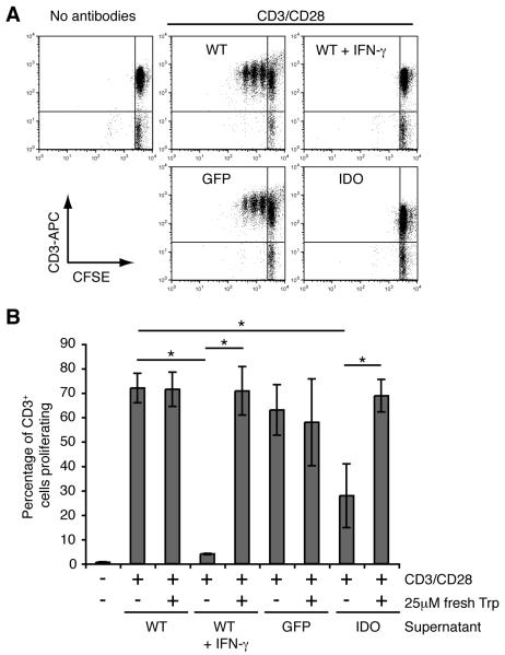
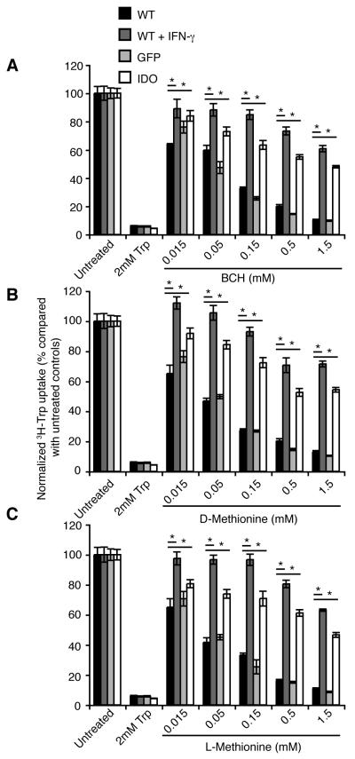
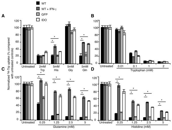
IDO is the rate-limiting enzyme in the kynurenine pathway, catabolizing tryptophan to kynurenine. Tryptophan depletion by IDO-expressing tumors is a common mechanism of immune evasion inducing regulatory T cells and inhibiting effector T cells. Because mammalian cells cannot synthesize tryptophan, it remains unclear how IDO(+) tumor cells overcome the detrimental effects of local tryptophan depletion. We demonstrate that IDO(+) tumor cells express a novel amino acid transporter, which accounts for ∼50% of the tryptophan uptake. The induced transporter is biochemically distinguished from the constitutively expressed tryptophan transporter System L by increased resistance to inhibitors of System L, resistance to inhibition by high concentrations of most amino acids tested, and high substrate specificity for tryptophan. Under conditions of low extracellular tryptophan, expression of this novel transporter significantly increases tryptophan entry into IDO(+) tumors relative to tryptophan uptake through the low-affinity System L alone, and further decreases tryptophan levels in the microenvironment. Targeting this additional tryptophan transporter could be a way of pharmacological inhibition of IDO-mediated tumor escape. These findings highlight the ability of IDO-expressing tumor cells to thrive in a tryptophan-depleted microenvironment by expressing a novel, highly tryptophan-specific transporter, which is resistant to inhibition by most other amino acids. The additional transporter allows tumor cells to strike the ideal balance between supply of tryptophan essential for their own proliferation and survival, and depleting the extracellular milieu of tryptophan to inhibit T cell proliferation.


Figures

Figure 1
Figure 1. IDO positive EG7 cells express an alternative BCH resistant tryptophan transporter in addition to System L
3H-Tryptophan uptake was measured over 3 minutes in EG7 WT, GFP-transduced or IDO-transduced (C7) cells after incubating for 30 minutes in sodium and amino acid free buffer. Uptake measurements were performed in duplicate and were measured in the presence of different concentrations of different inhibitors as indicated. A. Unlabelled tryptophan competes with the 3H-tryptophan for uptake through all tryptophan transporters and has a similar effect on tryptophan uptake by each cell type. B. BCH inhibits tryptophan uptake through System L, and is more potent at inhibiting uptake in IDO negative tumor cells. C. D-Met and D. the isomer L-Met inhibit tryptophan uptake through LAT1 mediated transporters and are more effective in IDO negative cells. Data are normalized to uptake in untreated cells and are shown as the means of duplicate samples from at least two combined independent experiments ± SE. Statistical significance <0.05 as assessed using Student’s T-test is indicated by *. Data are representative of at least three independent experiments.
Figure 2
Figure 2. Inhibition of T cell proliferation by IDO positive Hela cells by depletion of tryptophan
Peripheral blood lymphocytes were labeled with CFSE and cultured in preconditioned supernatants from Hela cells in 96 well flat-bottomed plates, pre-coated with PBS or with antibodies against human CD3 and CD28. After 4d the cells were harvested, stained with anti human CD3-APC and acquired on the flow cytometer (FACScalibur), gating on propidium iodide negative (live) cells and analyzed using FlowJo software. A. Representative dot plots are shown from cells cultured in conditioned supernatant from WT, WT + IFN-γ, GFP or IDO Hela cells, with CFSE against CD3-APC and T cell proliferation is shown by dilution of CFSE. B. T cell proliferation in pre-conditioned supernatant can be restored to normal levels by adding 25μM fresh L-tryptophan to the T cell cultures. Data are shown as percentage of CD3+ T cells that have undergone proliferation and are the means of duplicate wells from two independent experiments combined ± SE. The data shown are representative from at least three experiments. Statistical significance <0.05 as assessed using Student’s T-test is indicated by *.
Figure 3
Figure 3. Human tumor cells expressing either IDO through transfection or in response to IFN-γ express a BCH resistant tryptophan transporter
Measurement of transporter-mediated 3H-L-tryptophan uptake was measured under initial rate conditions over 3 min. Hela cells were cultured for 48h before the assay and tryptophan uptake by IDO-lentivirus transduced or IFN-γ treated Hela cells was measured in the presence of a panel of System L and LAT inhibitors including: A. BCH, B. D-Met and C. L-Met. Plotted data are normalized and are the means of triplicate measurements ± SE. Statistical significance <0.05 as assessed using Student’s T-test is indicated by *. Data are representative of at least three independent experiments.
Figure 4
Figure 4. Tryptophan uptake by IDO negative, but not IDO positive Hela cells is inhibited in the presence of glutamine or histidine
3H-Tryptophan uptake was measured in the presence of unlabeled amino acids to determine the amino acid specificity of the induced transporter in IDO positive cells. A. Tryptophan uptake was measured in the presence of high concentrations of a panel of unlabeled amino acids individually including L-tryptophan, histidine, glycine and glutamine. Tryptophan uptake was inhibited in all cell types tested equally by tryptophan, while glycine had no effect. Histidine or glutamine inhibited tryptophan uptake less effectively by IDO positive compared with IDO negative cells. Titration experiments were performed measuring tryptophan uptake in the presence of a range of concentrations of: B. tryptophan, C. glutamine or D. histidine, indicating equal inhibition by tryptophan across cell types while incomplete inhibition of tryptophan uptake in IDO positive Hela cells with either glutamine or histidine. Plotted data are normalized and the means of triplicate measurements ± SE and are representative of at least two independent experiments. Statistical significance <0.05 as assessed using Student’s T-test is indicated by *. Experiments were performed in 12 well plates.
Figure 5
Figure 5. 3H-Tryptophan uptake by IDO positive cells in the presence of unlabeled glutamine is inhibited by a range of aromatic amino acids
A. Unlabelled tryptophan in the presence of 2.5mM glutamine inhibits tryptophan uptake by IDO positive cells with a Ki of 2.3-3.2μM. Curve fitting in the presence of glutamine and tryptophan was performed using Sigmaplot. Individual data points are indicated by open (Hela WT) or filled (GFP) triangles or by open (WT + IFN-γ) or filled circles (IDO). The IDO induced tryptophan transporter is inhibited by some of the aromatic amino acids and their derivatives. B. tyrosine, C. phenylalanine and D. L-Dopa were tested in the presence of 2.5mM glutamine at a range of concentrations. Plotted data are normalized and the means of triplicate measurements ± SE and are representative of at least two independent experiments. Statistical significance <0.05 as assessed using Student’s T-test is indicated by *. Experiments were performed in 12 well plates.

References

    1. Brady FO. Inhibition of rabbit intestinal indoleamine 2,3-dioxygenase by copper chelators. FEBS Lett. 1975;57:237–240. - PubMed
    1. Brady FO. Tryptophan 2,3-dioxygenase: a review of the roles of the heme and copper cofactors in catalysis. Bioinorg Chem. 1975;5:167–182. - PubMed
    1. Hayaishi O, Hirata F, Fujiwara M, Senoh S, Tokuyama T. Indoleamine 2,3-dioxygenase. Note II. Biological function. Acta Vitaminol Enzymol. 1975;29:291–293. - PubMed
    1. Ozaki Y, Edelstein MP, Duch DS. The actions of interferon and antiinflammatory agents of induction of indoleamine 2,3-dioxygenase in human peripheral blood monocytes. Biochem Biophys Res Commun. 1987;144:1147–1153. - PubMed
    1. Yoshida R, Imanishi J, Oku T, Kishida T, Hayaishi O. Induction of pulmonary indoleamine 2,3-dioxygenase by interferon. Proc Natl Acad Sci USA. 1981;78:129–132. - PMC - PubMed

Publication types

Substances