. 2011 Jul;23(7):2725-37.
doi: 10.1105/tpc.111.088088. Epub 2011 Jul 8.

The FRD3 citrate effluxer promotes iron nutrition between symplastically disconnected tissues throughout Arabidopsis development

Affiliations

The FRD3 citrate effluxer promotes iron nutrition between symplastically disconnected tissues throughout Arabidopsis development

Hannetz Roschzttardtz et al. Plant Cell. 2011 Jul.

Abstract

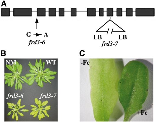
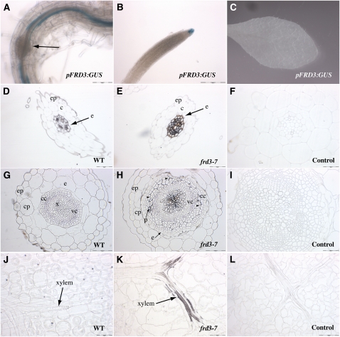
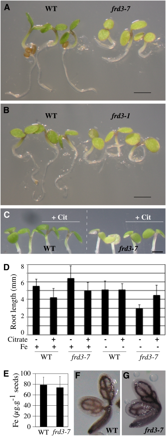
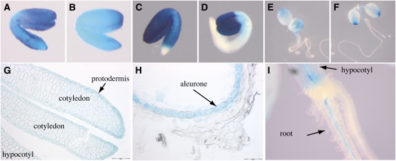
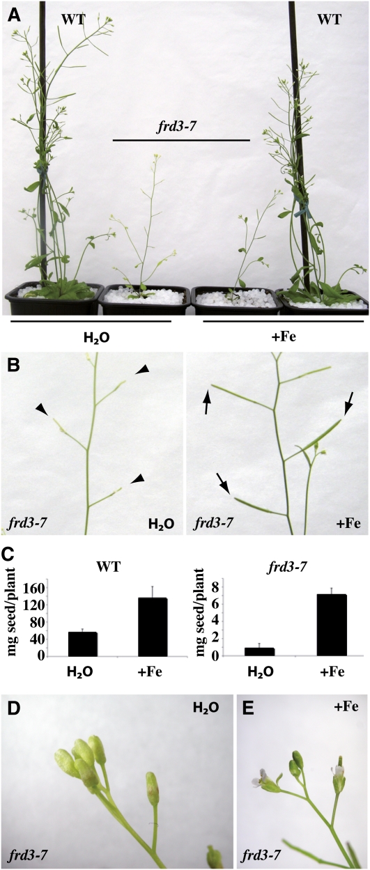
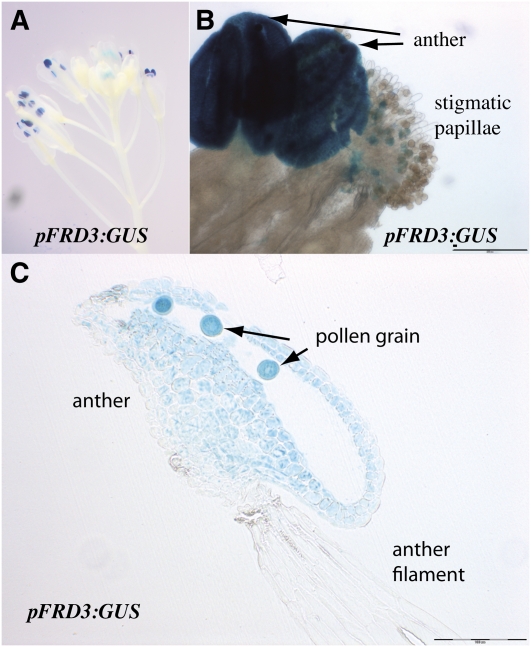
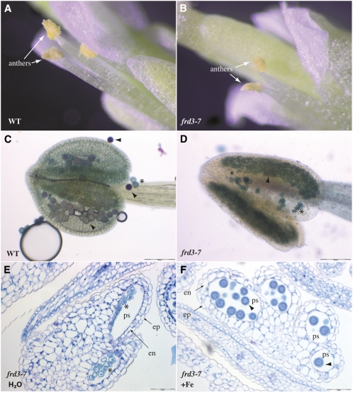
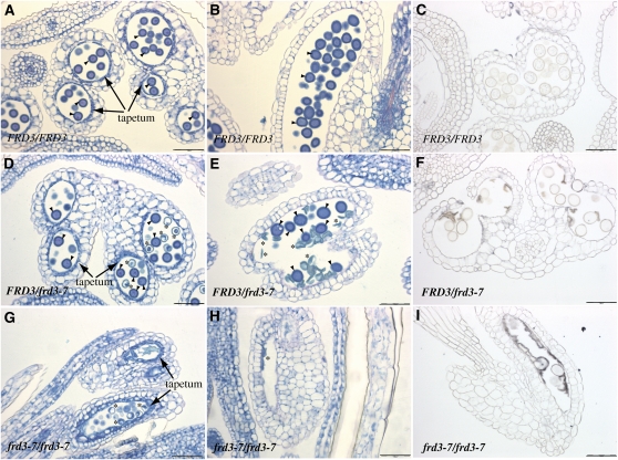
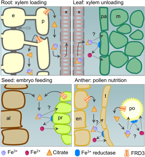
We present data supporting a general role for FERRIC REDICTASE DEFECTIVE3 (FRD3), an efflux transporter of the efficient iron chelator citrate, in maintaining iron homeostasis throughout plant development. In addition to its well-known expression in root, we show that FRD3 is strongly expressed in Arabidopsis thaliana seed and flower. Consistently, frd3 loss-of-function mutants are defective in early germination and are almost completely sterile, both defects being rescued by iron and/or citrate supply. The frd3 fertility defect is caused by pollen abortion and is associated with the male gametophytic expression of FRD3. Iron imaging shows the presence of important deposits of iron on the surface of aborted pollen grains. This points to a role for FRD3 and citrate in proper iron nutrition of embryo and pollen. Based on the findings that iron acquisition in embryo, leaf, and pollen depends on FRD3, we propose that FRD3 mediated-citrate release in the apoplastic space represents an important process by which efficient iron nutrition is achieved between adjacent tissues lacking symplastic connections. These results reveal a physiological role for citrate in the apoplastic transport of iron throughout development, and provide a general model for multicellular organisms in the cell-to-cell transport of iron involving extracellular circulation.


Figures

Figure 1.
Figure 1.
Phenotype of Two New Alleles of FRD3. (A) Position of frd3-6 and frd3-7 mutations on the FRD3 gene. LB, left border; G → A, guanine to adenine transversion. (B) Phenotype of 7-week-old frd3-6 and frd3-7 mutants grown in soil. NM, nonmutagenized parental line; WT, wild type. (C) frd3 chlorosis is reverted by iron supply. Four-week-old frd3-6 plants cultivated on soil were irrigated with water (−Fe) or 0.5 mM FeEDDHA (+Fe).
Figure 2.
Figure 2.
FRD3 Promoter Activity in Root and Shoot, and Iron Distribution in the frd3-7 Mutant. (A) to (C) Histochemical GUS staining of proFRD3:GUS plants from a representative transgenic line. (A) and (B) Primary root from a 1-week-old axenically grown plant in the mature zone (A) or the meristematic zone (B). The arrow in (A) shows a developing lateral root. (C) Rosette leaf of a 4-week-old soil-grown plant. (D) to (L) Perls/DAB staining of thin sections of 4-week-old wild-type (WT; [D], [G], and [J]) or frd3-7 plants ([E], [F], [H], [I], [K], and [L]). Roots ([D] to [F]), hypocotyl ([G] to [I]), and rosette leaves ([J] to [L]) are shown; (F), (I), and (L) are staining negative controls without Perls. c, cortex; cc, cork cambium; cp, cortex parenchyma; e, endodermis; ep, epidermis; p, phloem; vc, vascular cambium; x, xylem; arrowheads in (H) indicate the line of separation between vascular and cork cambium. Scale bars = 50 μm in (D), (E), (I), (J), (K), and (L), 100 μm in (A), (G), and (H), and 200 μm in (B).
Figure 3.
Figure 3.
FRD3 Is Required for Efficient Germination in Iron-Deficient Conditions. (A) and (B) Chlorosis and reduced size of 3-d-old frd3-7 (A) and frd3-1 (B) seedlings grown under iron deficiency (no added iron) compared with wild-type (WT) plants. (C) and (D) Citrate supplementation partially rescues the germination defect of the frd3 mutant. Wild-type and frd3-7 plants were grown for 3.5 d under iron sufficiency (50 μM FeEDTA, +Fe) or iron deficiency (no added iron) in the presence or absence of 3 mM Na citrate (+Cit) as indicated. Citrate treatment rescues frd3-7 chlorosis (C) and root growth defect (D). Scale bars = 1 mm. (E) Iron content in wild-type and frd3-7 seeds. Error bars = SD. (F) and (G) Iron localization in wild-type (F) and frd3-7 (G) dry seed embryos assayed by Perls/DAB histochemical staining.
Figure 4.
Figure 4.
The FRD3 Promoter Is Active during Embryogenesis and the Early Stages of Germination. GUS histochemical staining of a representative proFRD3:GUS transgenic line. (A) to (C) Dissected embryos from green siliques ([A] and [B]) or a dry seed (C). (D) to (F) Seedling after 1 d (D), 3 d (E), or 5 d (F) of germination. (G and H) Longitudinal section of a dry seed showing the embryo (G) or the seed envelope (H). (I) Close-up image of the hypocotyl-root transition zone of a 5-d-old seedling. Scale bars = 50 μm
Figure 5.
Figure 5.
Phenotype of the frd3 Mutant at the Reproductive Stage. Wild-type and frd3-7 plants were grown for 8 weeks in soil irrigated with water or 500 μM FeEDDHA. (A) to (E) Phenotypic defects of the frd3-7 mutant and their rescue by iron. Chlorosis (A), production of altered siliques (B), reduced seed production (C), and absence of flower opening ([D] and [E]). Elongated (normal) and nonelongated (mutant) siliques are labeled with arrows and arrowheads, respectively (B). The graph (C) represents the mean of two independent experiments, each performed with six to eight plants. Error bars = SD. WT, wild type. [See online article for color version of this figure.]
Figure 6.
Figure 6.
FRD3 Promoter Activity in Flower. GUS histochemical staining of an inflorescence of a representative pFRD3:GUS transgenic line. (A) Inflorescence. (B) Anther and stigmatic papillae. (C) Longitudinal section of a stamen. Scale bars = 100 μm.
Figure 7.
Figure 7.
Pollen Development Is Impaired in frd3-7. (A) and (B) Detailed view of a wild-type (WT; [A]) and an frd3-7 flower (B), highlighting the morphological difference of the frd3-7 anthers. (C) and (D) Pollen viability assay in wild-type (C) or frd3-7 anthers (D) using the Alexander’s stain. Viable and dead pollen grains are stained in pink and blue, respectively. (E) and (F) Periodic acid-Schiff-naphthol blue-black–stained cross sections of mature flowers collected from frd3-7 plants irrigated (F) or not (E) with 0.5 μM FeEDDHA. Arrowhead, normal pollen; asterisk, abnormal pollen; ep, epidermis; en, endothecium; ps, pollen sac. Scale bars = 50 μm in (E) and (F) and 100 μm in (C) and (D).
Figure 8.
Figure 8.
The frd3 Mutation Acts Gametophytically to Alter Pollen Development. (A) to (I) Histological sections of flowers from wild-type FRD3/FRD3 ([A] to [C]), heterozygous FRD3/frd3-7 ([D] to [F]), or homozygous mutant frd3-7/frd3-7 plants ([G] and [H]) stained with periodic acid-Schiff-naphthol blue-black ([A], [B], [D], [E], [G], and [H]) or with Perls/DAB ([C], [F], and [I]). (A), (D), and (G) Immature anther containing pollen at the two-cell stage. (B), (C), (E), (F), (H), and (I) Mature anther containing trinucleated pollen before dehiscence. Arrowhead, normal pollen; asterisk, abnormal pollen. Scale bars = 50 μm.
Figure 9.
Figure 9.
Model for the Role of FRD3 in Iron Nutrition between Symplastically Disconnected Tissues. Schematic representation of the proposed role of FRD3 in the different organs at different developmental stages. Top left, FRD3 in the root pericycle is required for solubilization of iron in the apoplast surrounding the stele cells and for proper loading of iron in the xylem sap. This citrate efflux activity might function in coordination with an iron efflux activity from endodermis or pericycle. Top right, Although not produced in leaves, citrate released by FRD3 in the central apoplast of the root is necessary for proper iron loading in the limb cells of the leaf. Bottom left, In the seed, where FRD3 expression is high in the embryo, particularly in the protodermis, and in the aleurone layer of the seed envelope, FRD3 might function to release citrate in the space separating the embryo and the envelope to provide available iron to feed the embryo. Bottom right, in the anther, the pollen grain depends on the pollen expression of FRD3 for its maturation and on the sporophytic expression of FRD3 for optimal ion loading, indicating that citrate efflux in the pollen sac is likely to increase iron solubility and to promote, directly or indirectly, iron acquisition by the pollen grain. In addition, tapetum morphogenesis requires a functional FRD3. In the three latter organs, seed, anther, and leaf, subsequent entry of iron in the sink tissue could occur as an Fe(III)-citrate complex by an as yet unidentified transporter or, after reduction into Fe(II), through a divalent metal transporter of the ZRT-IRT LIKE PROTEIN or Natural Resistance-Associated Macrophage Protein1 families. Because the four above-described situations share the common feature that iron transport from source to sink tissues involves an apoplastic step, we propose that FRD3-mediated citrate efflux is a prerequisite for iron traffic between symplastically disconnected tissues. al, aleurone; e, endodermis; en, endothecium; m, mesophyll cell; p, pericycle; pa, parenchyma; po, pollen grain; pr, protodermis; t, tapetum; x, xylem. [See online article for color version of this figure.]

Similar articles

Cited by

References

    1. Brown J.C. (1966). Fe and Ca uptake as related to root-sap and stem-exudate citrate in soybeans. Physiol. Plant. 19: 968–976
    1. Cailliatte R., Schikora A., Briat J.F., Mari S., Curie C. (2010). High-affinity manganese uptake by the metal transporter NRAMP1 is essential for Arabidopsis growth in low manganese conditions. Plant Cell 22: 904–917 - PMC - PubMed
    1. Clough S.J., Bent A.F. (1998). Floral dip: A simplified method for Agrobacterium-mediated transformation of Arabidopsis thaliana. Plant J. 16: 735–743 - PubMed
    1. Colangelo E.P., Guerinot M.L. (2004). The essential basic helix-loop-helix protein FIT1 is required for the iron deficiency response. Plant Cell 16: 3400–3412 - PMC - PubMed
    1. Connolly E.L., Fett J.P., Guerinot M.L. (2002). Expression of the IRT1 metal transporter is controlled by metals at the levels of transcript and protein accumulation. Plant Cell 14: 1347–1357 - PMC - PubMed

Publication types

MeSH terms