. 2011 Aug 22;52(9):6671-9.
doi: 10.1167/iovs.11-7559.

Heparan sulfate regulates intraretinal axon pathfinding by retinal ganglion cells

Affiliations

Heparan sulfate regulates intraretinal axon pathfinding by retinal ganglion cells

Minako Ogata-Iwao et al. Invest Ophthalmol Vis Sci. .

Abstract

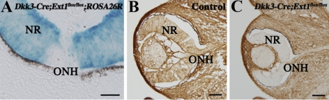
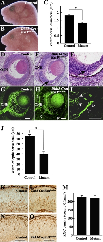
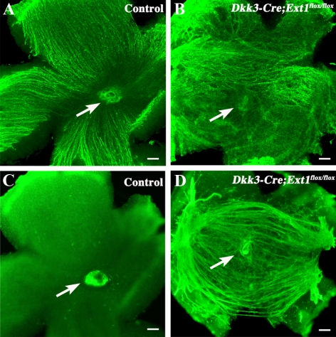
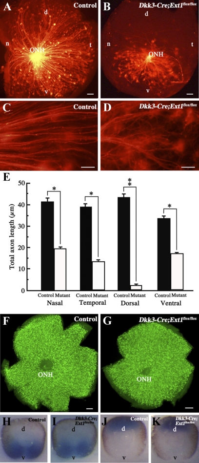
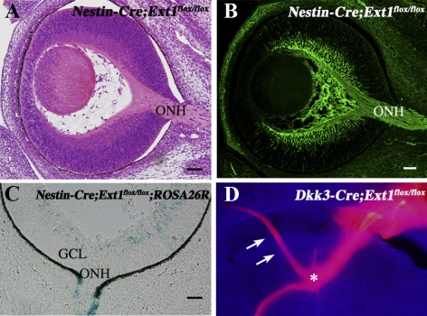
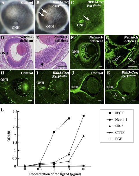
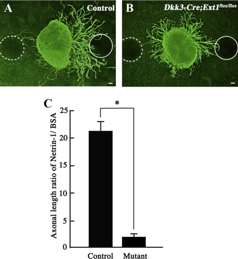
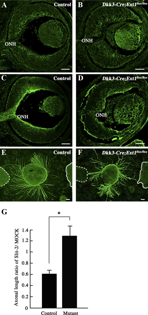
PURPOSE. Heparan sulfate (HS) is abundantly expressed in the developing neural retina; however, its role in the intraretinal axon guidance of retinal ganglion cells (RGCs) remains unclear. In this study, the authors examined whether HS was essential for the axon guidance of RGCs toward the optic nerve head. METHODS. The authors conditionally ablated the gene encoding the exostosin-1 (Ext1) enzyme, using the dickkopf homolog 3 (Dkk3)-Cre transgene, which disrupted HS expression in the mouse retina during directed pathfinding by RGC axons toward the optic nerve head. In situ hybridization, immunohistochemistry, DiI tracing, binding assay, and retinal explant assays were performed to evaluate the phenotypes of the mutants and the roles of HS in intraretinal axon guidance. RESULTS. Despite no gross abnormality in RGC distribution, the mutant RGC axons exhibited severe intraretinal guidance errors, including optic nerve hypoplasia, ectopic axon penetration through the full thickness of the neural retina and into the subretinal space, and disturbance of the centrifugal projection of RGC axons toward the optic nerve head. These abnormal phenotypes shared similarities with the RGC axon misguidance caused by mutations of genes encoding Netrin-1 and Slit-1/2. Explant assays revealed that the mutant RGCs exhibited disturbed Netrin-1-dependent axon outgrowth and Slit-2-dependent repulsion. CONCLUSIONS. The present study demonstrated that RGC axon projection toward the optic nerve head requires the expression of HS in the neural retina, suggesting that HS in the retina functions as an essential modulator of Netrin-1 and Slit-mediated intraretinal RGC axon guidance.


Figures

Figure 1.
Figure 1.
Disrupted expression of HS in the developing neural retina. (A) Dkk3-Cre-driven recombination in the developing neural retina. Frozen retinal sections of the Dkk3-Cre;Ext1flox/flox;ROSA26R mutants showed intense β-galactosidase activity throughout the neural retina at E11.5, indicating Cre-driven recombination before the optic nerve head formation. (B, C) Immunohistochemistry for HS in the control (B) and Dkk3-Cre;Ext1flox/flox (C) eyes at E12.5. Note that the HS immunoreactivity had disappeared in the mutant neural retina. NR, neural retina; ONH, optic nerve head. Scale bars, 50 μm.
Figure 2.
Figure 2.
Phenotypes of Dkk3-Cre;Ext1flox/flox embryos. (AC) Stereomicroscopy of control (A) and Dkk3-Cre;Ext1flox/flox (B) embryos at E18.5. The ventrodorsal diameters (C) of the Dkk3-Cre;Ext1flox/flox eyes were significantly smaller than those of the control eyes. Data represent mean ± SE. *P < 0.001, Wilcoxon signed-rank test (n = 5 for each). (DF) HE-stained sagittal sections of the control (D) and Dkk3-Cre;Ext1flox/flox (E, F) eyes at E14.5. (arrows) Ectopic axon bundles that penetrated the full thickness of the mutant peripheral retina. (GJ) Tuj1 immunohistochemistry in the control (G) and Dkk3-Cre;Ext1flox/flox (H, I) eyes at E14.5. Arrowheads (I) and open arrowheads (H, I) show ectopic axon bundles in the intraretinal region and the subretinal space, respectively. The width (J) of the Tuj1-positive optic nerve head of Dkk3-Cre;Ext1flox/flox mutants was significantly smaller than that of the controls. Data represent mean ± SE *P < 0.001, Wilcoxon signed-rank test (n = 5 for each). (KM) Immunohistochemistry for the RGC marker, Brn3, in sagittal sections of the control (K) and Dkk3-Cre;Ext1flox/flox (L) eyes at E14.5. There was no significant difference in RGC density between the controls and mutants (M). *P = 0.7369, Wilcoxon signed-rank test (n = 4 for each). (N, O) Immunohistochemistry for the photoreceptor cell marker Otx2 in sagittal sections of the control (N) and Dkk3-Cre;Ext1flox/flox (O) eyes at E14.5, demonstrating no difference in the distributions. Mutant, Dkk3-Cre;Ext1flox/flox; ONH, optic nerve head; Scale bars, 50 μm.
Figure 3.
Figure 3.
Tuj1 immunohistochemistry in flat-mounted retinas. (A, B) Vitreous side of the flat-mounted retinas of the control (A) and Dkk3-Cre;Ext1flox/flox (B) embryos at E16.5. Note the disturbed projection toward the optic nerve head in the mutant RGC axons. (C, D) Retinal pigment epithelium side of the flat-mounted retinas of the control (C) and Dkk3-Cre;Ext1flox/flox (D) embryos at E16.5. Numerous ectopic axons were present at the subretinal level of the mutant retina. (arrows) Optic nerve head. Scale bars, 50 μm.
Figure 4.
Figure 4.
Retrograde labeling of RGCs with axons extended toward the optic nerve head. (AE) Flat-mounted retinas after retrograde DiI labeling of the control (A, C) and Dkk3-Cre;Ext1flox/flox (B, D) embryos at E14.5. Fewer axon projections of RGCs toward the optic nerve head were observed in the mutant retina. This reduction was significant in each quadrant (E). Data represent mean ± SE. *P < 0.05, **P < 0.01, Wilcoxon signed-rank test (n = 5). The mutant axon projection was twisted (D) compared with the controls (C). (F, G) Immunohistochemistry for Brn3 in flat-mounted retina. Brn3-positive cells corresponding to RGCs were distributed throughout the retinas of the controls (F) and the mutants (G). (HK) Whole-mount in situ hybridization for Vax2 and Tbx5. There were no differences in the expression patterns of Vax2 or Tbx5 between the control (H, Vax2; J, Tbx5) and mutant (I, Vax2; K, Tbx5) retinas. d, dorsal; n, nasal; t, temporal; v, ventral; ONH, optic nerve head. Scale bars, 50 μm.
Figure 5.
Figure 5.
Retinal phenotypes in Nestin-Cre;Ext1flox/flox mutants. (A, B) HE-stained (A) and Tuj1-immunostained (B) sagittal retinal sections at E14.5 demonstrated no optic nerve head hypoplasia or ectopic penetration of RGC axons within the retina. (C) X-gal staining in the retinal sample of a Nestin-Cre;Ext1flox/flox;ROSA26R embryo at E14.5. Some retinal cells had β-galactosidase activity, but it was barely detectable in retinal samples up until E12.5, indicating the Cre-driven recombination that was late for the optic nerve formation. (D) Ectopic projection into the contralateral optic nerve at the Dkk3-Cre;Ext1flox/flox optic chiasm. DiI crystals were implanted onto the optic nerve head of the mutant right eye at E18.5. The anterogradely DiI-labeled optic nerve was misrouted into the optic nerve of the left eye (arrow) at the optic chiasm (asterisk); this phenotype was observed at the optic chiasm of Nestin-Cre;Ext1flox/flox mutants in a previous report. GCL, ganglion cell layer; ONH, optic nerve head. Scale bars, 50 μm.
Figure 6.
Figure 6.
Retinal phenotypes of Netrin-1–deficient and Dkk3-Cre;Ext1flox/flox mutants. (AC) Posterior views of the control (A) and Dkk3-Cre;Ext1flox/flox (B) eyes at E16.5. (arrows) Hypopigmented streaks in the retinal pigment epithelium. The streaks in the Dkk3-Cre;Ext1flox/flox eyes exhibited Tuj-1 immunoreactivity, suggestive of ectopic axon bundles from the retina (C). (D, E) HE-stained sagittal sections of the Netrin-1–deficient retina at E14.5. (D, E) Optic nerve head hypoplasia was observed. (E, arrowhead) Ectopic axon bundles. (F, G) Tuj1 immunohistochemistry in sagittal sections of Netrin-1–deficient retina at E14.5. Optic nerve head hypoplasia and ectopic axon bundles (open arrows), similar to the phenotypes of Dkk3-Cre;Ext1flox/flox mutants, were observed in Netrin-1–deficient mutants. (HK) Immunohistochemistry for netrin-1 and DCC. In the control retina, Netrin-1 (H) and DCC (J) immunoreactivities were observed around the optic nerve heads and the RGC axon bundles, respectively. In the Dkk3-Cre;Ext1flox/flox retina, netrin-1 (I) was also distributed around the hypoplastic optic nerve head. DCC (K) in the mutant retina was distributed in the RGC axon bundles and in the ectopic axon bundles (open arrowheads). (L) HS-binding assay. Netrin-1, bFGF, and Slit-2 bound to HS, whereas EGF and CNTF did not. ONH, optic nerve head. Scale bars, 50 μm.
Figure 7.
Figure 7.
Loss of Netrin-1–dependent outgrowth of RGC axons in HS-deficient retinal explants. (A) Control retinal explant at E13.5 after 24 hours in vitro. Axon outgrowth was significantly more robust on the Netrin-1–absorbed bead (solid circle) than on the BSA-absorbed bead (broken circle). (B) Dkk3-Cre;Ext1flox/flox retinal explant at E13.5 after 24 hours in vitro. The mutant axon outgrowth showed no apparent dependence on Netrin-1. (C) The ratio of the total lengths of the axons derived from each aspect was calculated, as indicated in the graph, using the value for the Netrin-1–soaked bead divided by that for the BSA-soaked bead for each explant. The mutant retinal explants showed significantly less dependence on Netrin-1 than the control explants (n = 29 for each; *P < 0.0001; Wilcoxon signed-rank test). Mutant, Dkk3-Cre;Ext1flox/flox. Scale bars, 50 μm.
Figure 8.
Figure 8.
Loss of the Slit-2–dependent repulsive effect of RGC axons in the HS-deficient retinal explants. (AD) Immunohistochemistry for Slit-2 and Robo-2. In the control retina, Slit-2 (A) and Robo-2 (C) immunoreactivities were observed in the inner region of the neural retina and in the RGC axon bundles including the optic nerve head, respectively. In the Dkk3-Cre;Ext1flox/flox retina, Slit-2 (B) was also distributed in the inner region of the neural retina. Robo-2 (D) in the mutant retina was distributed in the RGC axon bundles and the ectopic axon bundles. (EG) Slit-2–induced chemorepulsive assay for axon outgrowth from a retinal explant (48 hours in vitro at E13.5). Axon outgrowth from the control explant (E) was repelled in the Slit-2–transected 293T cell aggregate (solid line), whereas the Dkk3-Cre;Ext1flox/flox axons (F) showed no repulsion in the Slit-2–transfected cell aggregate (solid line). (broken lines) Mock-transfected 293T cell aggregates. Statistical analysis (G) confirmed significantly less dependence on Slit-2 in the mutant retinal explants than in the control explants (n = 21 for control and n = 24 for Dkk3-Cre;Ext1flox/flox; *P = 0.0002; Wilcoxon signed-rank test). Mutant, Dkk3-Cre;Ext1flox/flox; ONH, optic nerve head. Scale bars, 50 μm.

Similar articles

Cited by

References

    1. Harada T, Harada C, Parada LF. Molecular regulation of visual system development: more than meets the eye. Genes Dev. 2007;21:367–378 - PubMed
    1. Inatani M. Molecular mechanisms of optic axon guidance. Naturwissenschaften. 2005;92:549–561 - PubMed
    1. Deiner MS, Kennedy TE, Fazeli A, Serafini T, Tessier-Lavigne M, Sretavan DW. Netrin-1 and DCC mediate axon guidance locally at the optic disc: loss of function leads to optic nerve hypoplasia. Neuron. 1997;19:575–589 - PubMed
    1. Birgbauer E, Cowan CA, Sretavan DW, Henkemeyer M. Kinase independent function of EphB receptors in retinal axon pathfinding to the optic disc from dorsal but not ventral retina. Development. 2000;127:1231–1241 - PubMed
    1. Bastmeyer M, Ott H, Leppert CA, Stuermer CA. Fish E587 glycoprotein, a member of the L1 family of cell adhesion molecules, participates in axonal fasciculation and the age-related order of ganglion cell axons in the goldfish retina. J Cell Biol. 1995;130:969–976 - PMC - PubMed

MeSH terms