. 2011 Aug 3;103(15):1190-204.
doi: 10.1093/jnci/djr243. Epub 2011 Jul 8.

Modulation of pancreatic cancer chemoresistance by inhibition of TAK1

Affiliations

Modulation of pancreatic cancer chemoresistance by inhibition of TAK1

Davide Melisi et al. J Natl Cancer Inst. .

Erratum in

Abstract

Background: TGF-β-activated kinase-1 (TAK1), a mitogen-activated protein kinase kinase kinase, functions in the activation of nuclear factor κB (NF-κB) and activator protein-1, which can suppress proapoptotic signaling pathways and thus promote resistance to chemotherapeutic drugs. However, it is not known if inhibition of TAK1 is effective in reducing chemoresistance to therapeutic drugs against pancreatic cancer.

Methods: NF-κB activity was measured by luciferase reporter assay in human pancreatic cancer cell lines AsPc-1, PANC-1, and MDAPanc-28, in which TAK1 expression was silenced by small hairpin RNA. TAK1 kinase activity was targeted in AsPc-1, PANC-1, MDAPanc-28, and Colo357FG cells with exposure to increasing doses of a selective small-molecule inhibitor, LYTAK1, for 24 hours. To test the effect of LYTAK1 in combination with chemotherapeutic agents, AsPc-1, PANC-1, MDAPanc-28 cells, and control cells were treated with increasing doses of oxaliplatin, SN-38, or gemcitabine in combination with LYTAK1. In vivo activity of oral LYTAK1 was evaluated in an orthotopic nude mouse model (n = 40, 5 per group) with luciferase-expressing AsPc-1 pancreatic cancer cells. The results of in vitro proliferation were analyzed for statistical significance of differences by nonlinear regression analysis; differences in mouse survival were determined using a log-rank test. All statistical tests were two-sided.

Results: AsPc-1 and MDAPanc-28 TAK1 knockdown cells had a statistically significantly lower NF-κB activity than did their respective control cell lines (relative luciferase activity: AsPc-1, mean = 0.18, 95% confidence interval [CI] = 0.10 to 0.27; control, mean = 3.06, 95% CI = 2.31 to 3.80; MDAPanc-28, mean = 0.30, 95% CI = 0.13 to 0.46; control, mean = 4.53, 95% CI = 3.43 to 5.63; both P < .001). TAK1 inhibitor LYTAK1 had potent in vitro cytotoxic activity in AsPc-1, PANC-1, MDAPanc-28, and Colo357FG cells, with IC(50) between 5 and 40 nM. LYTAK1 also potentiated the cytotoxicity of chemotherapeutic agents oxaliplatin, SN-38, and gemcitabine in AsPc-1, PANC-1, and MDAPanc-28 cells compared with control cells (P < .001). In nude mice, oral administration of LYTAK1 plus gemcitabine statistically significantly reduced tumor burden (gemcitabine vs gemcitabine plus LYTAK1, P = .03) and prolonged survival duration (median survival: gemcitabine, 82 days vs gemcitabine plus LYTAK1, 122 days; hazard ratio = 0.334, 95% CI = 0.027 to 0.826, P = .029).

Conclusions: The results of this study suggest that genetic silencing or inhibition of TAK1 kinase activity in vivo is a potential therapeutic approach to reversal of the intrinsic chemoresistance of pancreatic cancer.


Figures
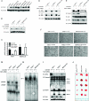
Figure 1
Figure 1
Determination of in vitro and in vivo proapoptotic phenotype by silencing TGF-β-activated kinase-1 (TAK1) expression. A) Western blot analysis for the expression of TAK1 in an immortalized and nontumorigenic human pancreatic ductal epithelial (HPDE) and eight different pancreatic cancer cell lines as indicated. B) Western blot analysis for the expression of TAK1 in AsPc-1, PANC-1, and MDAPanc-28 pancreatic cancer cells transduced with lentiviruses expressing TAK1-specific small hairpin RNA (shRNA) or a scramble sequence as control. C) TAK1 shRNA rescue experiment. Nuclear factor κB (NF-κB) reporter gene assay in AsPc-1 and MDAPanc-28 cells transduced with lentiviruses expressing TAK1-specific shRNA, a scramble sequence, or stably coexpressing human TAK1 shRNA and wild-type murine TAK1 sequences. The luciferase activities were normalized to the Renilla luciferase activity of the internal control. Error bars indicate 95% confidence intervals of two independent experiments performed in triplicate. Asterisks indicate statistical significance (P < .001) compared with scramble, as determined by two-sided one-way analysis of variance and Dunnett test. D) Western blot analysis for the autophosphorylation of TAK1 and E) the phosphorylation of p38 in AsPc-1, PANC-1, and MDAPanc-28 pancreatic cancer cells transduced with lentiviruses expressing TAK1-specific shRNA or a scramble sequence as control. F) Light-microscopic phenotype in AsPc-1, PANC-1, and MDAPanc-28 pancreatic cancer cells transduced with lentiviruses expressing TAK1-specific shRNA or a scramble sequence as control. Scale bars = 100 μm. G) Electrophoretic Mobility Shift Assay analysis for the NF-κB p65/p50 and p50/p50 complexes and H) activator protein-1 (AP-1) DNA-binding activity in AsPc-1, PANC-1, and MDAPanc-28 pancreatic cancer cells transduced with lentiviruses expressing TAK1-specific shRNA or a scramble sequence as control. I) Western blot analysis for the expression of cellular inhibitor of apoptosis 2 (cIAP-2), caspase-3, PARP-1, TAK1, and β-actin in the same cells as indicated. J) Fifteen athymic mice bearing orthotopic AsPc-1, AsPc-1scramble, or AsPc-1TAK1shRNA pancreatic tumors (n = 5 per group) were euthanized by carbon dioxide inhalation at the median survival duration of the control group (63 days). A digital image of resected tumors was acquired to assess tumor growth.
Figure 2
Figure 2
Effects of TGF-β-activated kinase-1 (TAK1) silencing on chemoresistance of pancreatic cancer cells. Percent survival of AsPC-1, PANC-1, and MDAPanc-28 pancreatic cancer cells after treatment with increasing molar concentrations (log scale) of gemcitabine, SN-38, and oxaliplatin. Dimethyl sulfoxide–treated cells were assigned a value of 100% and designated as control. Means and 95% confidence intervals of three independent experiments performed in quadruplicate are shown. Curves were fitted by nonlinear regression analysis. shRNA = small hairpin RNA.
Figure 3
Figure 3
Nuclear factor κB (NF-κB) activation after treatment with cytotoxic agents. AsPc-1, PANC-1, and MDAPanc-28 pancreatic cancer cells transduced with lentiviruses expressing TGF-β-activated kinase-1 (TAK1)-specific small hairpin RNA (shRNA), or a scramble sequence as control were treated for 24 hours with doses of gemcitabine, SN-38, or oxaliplatin corresponding with the IC50 for the control cells or saline as control. A) Electrophoretic Mobility Shift Assay analysis was performed to determine the DNA-binding activity of NF-κB p65/p50 and p50/p50 complexes. B) Apoptosis was measured by western blot analysis for PARP-1 cleavage and DNA fragmentation detected by agarose gel electrophoresis. Similar results were obtained with three independent experiments.
Figure 4
Figure 4
Activity of the TGF-β-activated kinase-1 (TAK1) kinase–selective inhibitor LYTAK1 in vitro. A) Percent survival of AsPc-1, PANC-1, MDAPanc-28, and COLO357FG pancreatic cancer cells after treatment with increasing molar concentrations (log scale) of LYTAK1. Dimethyl sulfoxide (DMSO)–treated cells were assigned a value of 100% and designated as control. Means and 95% confidence intervals of three independent experiments performed in quadruplicate are shown. Curves were fitted by nonlinear regression analysis. B) Western blot analysis was performed to determine the expression and phosphorylation of TAK1 and p38, and C) Electrophoretic Mobility Shift Assay analysis was performed to determine the DNA-binding activity of NF-κB p65/p50 and p50/p50 complexes in AsPc-1, PANC-1, and MDAPanc-28 pancreatic cancer cells treated for 24 hours with increasing doses of LYTAK1 or DMSO as control. Similar results were obtained with three independent experiments.
Figure 5
Figure 5
Inhibition of TGF-β-activated kinase-1 kinase activity and chemosensitization of human pancreatic cancer cells in vitro. Percent survival of AsPC-1, PANC-1, and MDAPanc-28 pancreatic cancer cell lines, and human papillomavirus type 16 early gene 6- and 7-immortalized/nontumorigenic human pancreatic ductal epithelial (HPDE) control cells after treatment with increasing molar concentrations (log scale) of gemcitabine, SN-38, and oxaliplatin alone or in combination with LYTAK1 at doses corresponding with the IC50 measured for each cell line or dimethyl sulfoxide (DMSO) as control. DMSO-treated cells were assigned a value of 100% and designated as control. Means and 95% confidence intervals of three independent experiments performed in quadruplicate are shown. Curves were fitted by nonlinear regression analysis.
Figure 6
Figure 6
LYTAK1 pharmacokinetic and pharmacodynamic models. BALB/c mice were treated (n = 3 per group) either with A) increasing doses of LYTAK1 for 2 hours or with or B) 6.9 mg/kg of LYTAK1 for different time points as indicated. Mouse blood was stimulated with tumor necrosis factor α (TNF-α), and TAK1 activity in peripheral blood mononuclear cells (Ly-6G−/CD11b+) was determined by flow cytometry with intracellular staining of p38 phosphorylation. Blue bars indicate mean fluorescence intensities (MFI) for phosphorylated p38 (p-p38). Error bars are SEs. Asterisks indicate P < .001 significance level compared with TNF-α-stimulated LYTAK1-untreated control, as determined by two-sided one-way analysis of variance and Dunnett test. Percentages indicate p-p38 MFI reduction compared with TNF-α-stimulated LYTAK1-untreated control. Red curves indicated drug exposure concentrations. Error bars are SEs.
Figure 7
Figure 7
Antitumor activity of oral LYTAK1 plus chemotherapeutic agents in vivo in AsPc-1 pancreatic tumor orthotopic xenografts (n = 40, 5 mice per group). A) Tumor volume was quantified as the sum of all detected photons within the region of the tumor per second. Error bars are 95% CI. *P = .0317, gemcitabine vs LYTAK1 plus gemcitabine by two-sided Mann–Whitney test. B) A digital grayscale image of each mouse was acquired, which was followed by the acquisition and overlay of a pseudocolor image representing the spatial distribution of detected photons emerging from active luciferase within the mouse. C) Mice were killed by carbon dioxide inhalation when evidence of advanced bulky disease was present. Survival was estimated from the day of pancreatic cancer cells orthotopic injection until the day of death. Differences among survival duration of mice in each group were determined by log-rank test. D) Immunohistochemical analysis. Serial paraffin sections from AsPc-1 tumors treated as indicated were stained with antibodies to the nuclear factor κB (NF-κB)– and activator protein-1–induced protein cellular inhibitor of apoptosis 2 (cIAP-2), the p65 subunit of NF-κB, and the NF-κB-induced protein interleukin 6 (IL-6).

Comment in

Similar articles

Cited by

References

    1. Jemal A, Siegel R, Xu J, Ward E. Cancer statistics, 2010. CA Cancer J Clin. 2010;60(5):277–300. - PubMed
    1. Melisi D, Chiao PJ. NF-kappa B as a target for cancer therapy. Expert Opin Ther Targets. 2007;11(2):133–144. - PubMed
    1. Verde P, Casalino L, Talotta F, Yaniv M, Weitzman JB. Deciphering AP-1 function in tumorigenesis: fra-ternizing on target promoters. Cell Cycle. 2007;6(21):2633–2639. - PubMed
    1. Rayet B, Gelinas C. Aberrant rel/nfkb genes and activity in human cancer. Oncogene. 1999;18(49):6938–6947. - PubMed
    1. Baldwin AS. Control of oncogenesis and cancer therapy resistance by the transcription factor NF-kappaB. J Clin Invest. 2001;107(3):241–246. - PMC - PubMed

Publication types

MeSH terms