Relationship between oral cancer and implants: clinical cases and systematic literature review
- PMID: 21743414
- PMCID: PMC3448182
- DOI: 10.4317/medoral.17223
Relationship between oral cancer and implants: clinical cases and systematic literature review
Abstract
The use of implants for oral rehabilitation of edentulous spaces has recently been on the increase, which has also led to an increase in complications such as peri-implant inflammation or peri-implantitis. Chronic inflammation is a risk factor for developing oral squamous cell carcinoma (OSCC).
Objectives: To review the literature of cases that associate implant placement with the development of oral cancer.
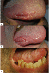
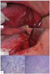
Study design: We present two clinical cases and a systematic review of literature published on the relationship between oral cancer and implants.
Results: We found 13 articles published between the years 1996 and 2009, referencing 18 cases in which the osseointegrated implants are associated with oral squamous cell carcinoma. Of those, 6 articles were excluded because they did not meet the inclusion criteria. Of the 18 cases reported, only 7 cases did not present a previous history of oral cancer or cancer in other parts of the body.
Conclusions: Based on the review of these cases, a clear cause-effect relationship cannot be established, although it can be deduced that there is a possibility that implant treatment may constitute an irritant and/or inflammatory cofactor which contributes to the formation and/or development of OSCC.
Figures


Similar articles
-
Interventions for replacing missing teeth: treatment of peri-implantitis.Cochrane Database Syst Rev. 2012 Jan 18;1(1):CD004970. doi: 10.1002/14651858.CD004970.pub5. Cochrane Database Syst Rev. 2012. PMID: 22258958 Free PMC article.
-
Sun protection for preventing basal cell and squamous cell skin cancers.Cochrane Database Syst Rev. 2016 Jul 25;7(7):CD011161. doi: 10.1002/14651858.CD011161.pub2. Cochrane Database Syst Rev. 2016. PMID: 27455163 Free PMC article.
-
Interventions for replacing missing teeth: management of soft tissues for dental implants.Cochrane Database Syst Rev. 2012 Feb 15;2012(2):CD006697. doi: 10.1002/14651858.CD006697.pub2. Cochrane Database Syst Rev. 2012. PMID: 22336822 Free PMC article.
-
Signs and symptoms to determine if a patient presenting in primary care or hospital outpatient settings has COVID-19.Cochrane Database Syst Rev. 2022 May 20;5(5):CD013665. doi: 10.1002/14651858.CD013665.pub3. Cochrane Database Syst Rev. 2022. PMID: 35593186 Free PMC article.
-
Soft tissue management for dental implants: what are the most effective techniques? A Cochrane systematic review.Eur J Oral Implantol. 2012 Autumn;5(3):221-38. Eur J Oral Implantol. 2012. PMID: 23000707
Cited by
-
Malignancy prediction among tissues from Oral SCC patients including neck invasions: a 1H HRMAS NMR based metabolomic study.Metabolomics. 2020 Mar 11;16(3):38. doi: 10.1007/s11306-020-01660-8. Metabolomics. 2020. PMID: 32162079
-
Oral Cancer around Dental Implants Appearing in Patients with\without a History of Oral or Systemic Malignancy: a Systematic Review.J Oral Maxillofac Res. 2017 Sep 30;8(3):e1. doi: 10.5037/jomr.2017.8301. eCollection 2017 Jul-Sep. J Oral Maxillofac Res. 2017. PMID: 29142653 Free PMC article. Review.
-
Novel Strategies for the Bioavailability Augmentation and Efficacy Improvement of Natural Products in Oral Cancer.Cancers (Basel). 2022 Dec 30;15(1):268. doi: 10.3390/cancers15010268. Cancers (Basel). 2022. PMID: 36612264 Free PMC article. Review.
-
An Update on Implant-Associated Malignancies and Their Biocompatibility.Int J Mol Sci. 2024 Apr 24;25(9):4653. doi: 10.3390/ijms25094653. Int J Mol Sci. 2024. PMID: 38731871 Free PMC article. Review.
-
Oral Lichen Planus and Dental Implants: Protocol and Systematic Review.J Clin Med. 2020 Dec 21;9(12):4127. doi: 10.3390/jcm9124127. J Clin Med. 2020. PMID: 33371347 Free PMC article. Review.
References
-
- Kademani D. Oral cancer. Mayo Clin Proc. 2007;82:878–887. - PubMed
-
- Kademani D, Bell RB, Schmidt BL, Blanchaert R, Fernandes R, Lambert P. Oral and maxillofacial surgeons treating oral cancer: a preliminaryreport from the American Association of Oral and Maxillofacial Surgeons TaskForce on Oral Cancer. J Oral Maxillofac Surg. 2008;66:2151–2157. - PubMed
-
- Warnakulasuriya KA, Ralhan R. Clinical, pathological, cellular and molecular lesions caused by oral smokeless tobacco--a review. J Oral Pathol Med. 2007;36:63–77. - PubMed
-
- Jane C, Nerurkar AV, Shirsat NV, Deshpande RB, Amrapurkar AD, Karjodkar FR. Increased survivin expression in high-grade oral squamous cell carcinoma: a study in Indian tobacco chewers. J Oral Pathol Med. 2006;35:595–601. - PubMed
-
- Guha N, Boffetta P, Wünsch Filho V, Eluf Neto J, Shangina O, Zaridze D. Oral health and risk of squamouscell carcinoma of the head and neck and esophagus: results of two multicentriccase-control studies. Am J Epidemiol. 2007;166:1159–1173. - PubMed

