. 2011 Jul 8;30(16):3309-21.
doi: 10.1038/emboj.2011.226.

PICH and BLM limit histone association with anaphase centromeric DNA threads and promote their resolution

Affiliations

PICH and BLM limit histone association with anaphase centromeric DNA threads and promote their resolution

Yuwen Ke et al. EMBO J. .

Abstract

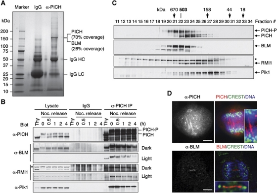
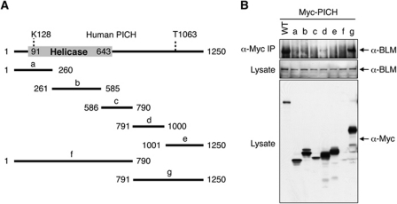
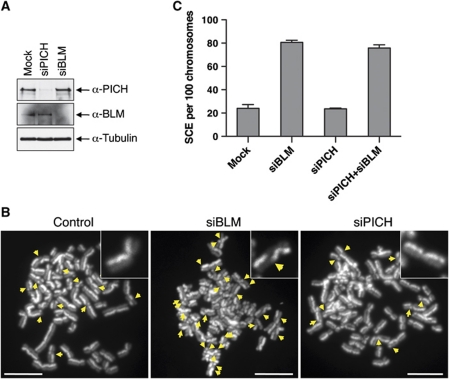
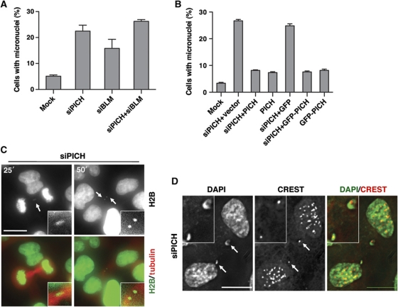
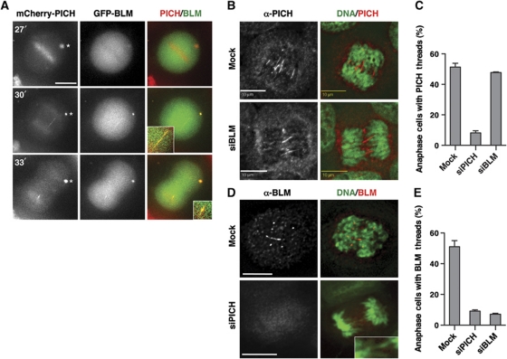
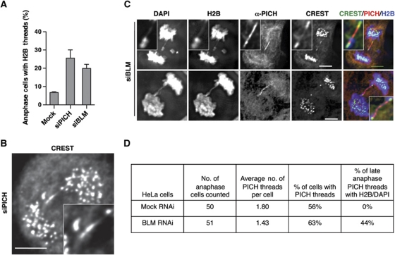
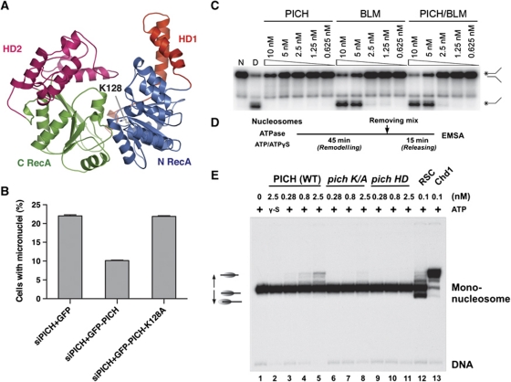
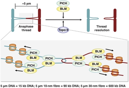
Centromeres nucleate the formation of kinetochores and are vital for chromosome segregation during mitosis. The SNF2 family helicase PICH (Plk1-interacting checkpoint helicase) and the BLM (the Bloom's syndrome protein) helicase decorate ultrafine histone-negative DNA threads that link the segregating sister centromeres during anaphase. The functions of PICH and BLM at these threads are not understood, however. Here, we show that PICH binds to BLM and enables BLM localization to anaphase centromeric threads. PICH- or BLM-RNAi cells fail to resolve these threads in anaphase. The fragmented threads form centromeric-chromatin-containing micronuclei in daughter cells. Anaphase threads in PICH- and BLM-RNAi cells contain histones and centromere markers. Recombinant purified PICH has nucleosome remodelling activities in vitro. We propose that PICH and BLM unravel centromeric chromatin and keep anaphase DNA threads mostly free of nucleosomes, thus allowing these threads to span long distances between rapidly segregating centromeres without breakage and providing a spatiotemporal window for their resolution.


Conflict of interest statement

The authors declare that they have no conflict of interest.

Figures

Figure 1
Figure 1
Identification of BLM as a PICH-binding protein. (A) Control IgG and anti-PICH IP from HeLa Tet-On cells were resolved by SDS–PAGE and stained with colloidal Coomassie blue. The molecular mass of markers is labelled. The sequence coverage of PICH and BLM in mass spectrometry is indicated. (B) Lysates, control IgG IP, or anti-PICH IP of HeLa Tet-On cells arrested in G1/S by thymidine (Thy) or at different times (hours) following the release from nocodazole-triggered mitotic arrest were blotted with the indicated antibodies. A cross-reacting band in the α-RMI1 blot is indicated with an asterisk. The positions of hyperphosphorylated and hypophosphorylated PICH are labelled. Light exposures of the α-BLM and α-RMI1 blots are included to better reveal their decrease in PICH binding during mitotic exit. (C) Lysates of nocodazole-arrested mitotic HeLa cells were fractionated on a Superose 6 column. The fractions were analysed by SDS–PAGE followed by immunoblotting with the indicated antibodies. The positions of hypophosphorylated and hyperphosphorylated forms of PICH are labelled. The faster migrating band in the α-BLM blot is derived from BLM, as it was depleted by BLM siRNA. A cross-reacting band in the α-RMI1 blot is indicated with an asterisk. The elution positions of the molecular mass standards are indicated. (D) HeLa Tet-On cells in anaphase were stained with anti-PICH or anti-BLM along with CREST (a centromere marker) and DAPI (stains DNA). In the overlay images, anti-PICH/BLM staining is in red. CREST staining is in green. DAPI staining is in blue. The PICH and BLM threads are magnified in the insets. Scale bars, 5 μm.
Figure 2
Figure 2
The C-terminal region of PICH binds to BLM. (A) Schematic drawing of the domain structure of human PICH. K128 is critical for ATP binding and for the helicase activity of PICH. Phosphorylation of T1063 creates a docking site for Plk1. The boundaries of the PICH truncation mutants are shown. (B) HeLa Tet-On cells were transfected with plasmids encoding either wild-type (WT) Myc-PICH or Myc-PICH truncation mutants described in (A). Lysates and anti-Myc IP of these cells were blotted with the indicated antibodies.
Figure 3
Figure 3
PICH is not required for suppressing SCE. (A) Lysates of HeLa Tet-On cells mock transfected or transfected with siPICH or siBLM were blotted with the indicated antibodies. (B) Representative metaphase spreads from the SCE assays of mock RNAi, BLM-RNAi, and PICH-RNAi HeLa Tet-On cells. The SCE events are indicated with arrows. A sister-chromatid pair from each metaphase spread is magnified and shown in insets. Scale bars, 10 μm. (C) Quantification of the number of SCE events per 100 chromosomes in cells either mock transfected or transfected with siBLM, siPICH, or both. Mean and s.d. of three independent experiments are shown.
Figure 4
Figure 4
PICH prevents formation of centromere-positive micronuclei. (A) Quantification of the percentage of micronuclei-containing HeLa Tet-On cells either mock transfected or transfected with siPICH, siBLM, or both. Mean and s.d. of three independent experiments are shown. (B) Ectopic expression of untagged PICH or GFP-PICH rescues the micronuclei phenotype of PICH-RNAi cells. Quantification of the percentage of micronuclei-containing HeLa Tet-On cells mock transfected or transfected with siPICH together with empty vector or plasmids encoding GFP or siPICH4-resistant PICH or GFP-PICH. Mean and s.d. of three independent experiments are shown. (C) Live cell imaging of PICH-RNAi HeLa Tet-On cells expressing H2B-mCherry (pseudo-coloured green) and GFP-tubulin (coloured red). Images at 25 and 60 min after NEBD are shown. Images from additional time points are shown in Supplementary Figure S6B. The H2B thread and micronuclei are magnified and shown in insets. Scale bar, 10 μm. (D) Representative images of PICH-RNAi cells stained with DAPI (green in overlay) and CREST (red in overlay). Two CREST-positive micronuclei are magnified and shown in insets. Scale bars, 10 μm.
Figure 5
Figure 5
PICH is required for BLM localization to anaphase threads. (A) Live cell imaging of HeLa Tet-On cells transfected with plasmids encoding mCherry-PICH (red in overlay) and GFP-BLM (green). Images at 27, 30, and 33 min after NEBD are shown. The PICH/BLM-positive thread and foci are magnified and shown in insets. Note that overexpressed PICH tended to form cytoplasmic foci (indicated with an asterisk), which also contained BLM at the 30- and 33-min time points. Scale bar, 10 μm. (B) Mock-transfected or BLM-RNAi HeLa Tet-On cells were stained with anti-PICH (red in overlay) and DAPI (green in overlay). Scale bars, 10 μm. (C) Quantification of the percentage of anaphase cells transfected with the indicated siRNAs that contained PICH threads. Mean and s.d. of three independent experiments are shown. (D) Mock-transfected or PICH-RNAi HeLa Tet-On cells were stained with anti-BLM (red in overlay) and DAPI (green in overlay). A DAPI-positive anaphase thread is shown in inset. Scale bars, 10 μm. (E) Quantification of the percentage of anaphase cells transfected with the indicated siRNAs that contained BLM threads. Mean and s.d. of three independent experiments are shown.
Figure 6
Figure 6
PICH and BLM prevent histone association with anaphase threads. (A) Quantification of the percentage of H2B-thread-containing anaphase HeLa Tet-On cells stably expressing H2B-GFP transfected with the indicated siRNAs. (B) A representative anaphase PICH-RNAi cell stained with CREST. An anaphase thread decorated with CREST is magnified and shown in inset. Scale bar, 5 μm. (C) Two representative anaphase BLM-RNAi H2B-GFP-expressing cells stained with DAPI, anti-PICH, and CREST. CREST, PICH, and H2B signals in the overlay are coloured green, red, and blue, respectively. PICH threads are magnified and shown in insets. Scale bars, 5 μm. (D) Statistics of the number of PICH threads and the percentage of PICH threads with histone H2B and DAPI in mock or BLM-RNAi anaphase HeLa cells.
Figure 7
Figure 7
BLM is required for anaphase thread resolution. (A) Quantification of SCE in PSNG13 (BLM−) and PSNF5 (BLM+) cells. (B) Statistics of the number of all PICH threads and the number of DAPI-positive PICH threads in anaphase PSNG13 (BLM−) and PSNF5 (BLM+) cells. (C) Representative images of anaphase PSNG13 (BLM−) and PSNF5 (BLM+) cells. The PICH thread in the PSNG13 cell contained DAPI while the PICH thread in the PSNF5 cell did not. Scale bars, 5 μm.
Figure 8
Figure 8
PICH has nucleosome sliding activity. (A) Ribbon diagram of a homology-based structure model of the HD of PICH. The model is built with the structure of Rad54 (PDB ID: 1Z3I) as the template. The ATP-binding residue K128 is shown in sticks. (B) Quantification of the percentage of micronuclei-containing HeLa Tet-On cells transfected with siPICH4 together with plasmids encoding GFP, siPICH4-resistant GFP-PICH, or GFP-PICH K128A. Mean and s.d. of three independent experiments are shown. (C) Helicase assay with PICH, BLM, and the PICH–BLM complex. Native (N) replication-fork-like duplex DNA and denatured (D) single-stranded DNA were included as controls. (D) Reaction schemes of the nucleosome sliding assay. (E) Nucleosome sliding assay of wild-type (WT) PICH, PICH K128A (K/A), PICH HD, RSC, and Chd1.
Figure 9
Figure 9
Model of PICH/BLM-dependent resolution of anaphase centromeric DNA threads. In this model, PICH and BLM collaborate to keep anaphase DNA threads largely free of nucleosomes, providing a spatiotemporal window for Topo II and possibly other DNA repair enzymes to resolve catenated DNA or other aberrant DNA structures.

Similar articles

Cited by

References

    1. Bachrati CZ, Borts RH, Hickson ID (2006) Mobile D-loops are a preferred substrate for the Bloom's syndrome helicase. Nucleic Acids Res 34: 2269–2279 - PMC - PubMed
    1. Baumann C, Korner R, Hofmann K, Nigg EA (2007) PICH, a centromere-associated SNF2 family ATPase, is regulated by Plk1 and required for the spindle checkpoint. Cell 128: 101–114 - PubMed
    1. Bharadwaj R, Yu H (2004) The spindle checkpoint, aneuploidy, and cancer. Oncogene 23: 2016–2027 - PubMed
    1. Bugreev DV, Yu X, Egelman EH, Mazin AV (2007) Novel pro- and anti-recombination activities of the Bloom's syndrome helicase. Genes Dev 21: 3085–3094 - PMC - PubMed
    1. Buscaino A, Allshire R, Pidoux A (2010) Building centromeres: home sweet home or a nomadic existence? Curr Opin Genet Dev 20: 118–126 - PubMed

Publication types