. 2011 Jul 10;17(8):1010-4.
doi: 10.1038/nm.2409.

Imaging the subcellular structure of human coronary atherosclerosis using micro-optical coherence tomography

Affiliations

Imaging the subcellular structure of human coronary atherosclerosis using micro-optical coherence tomography

Linbo Liu et al. Nat Med. .

Abstract

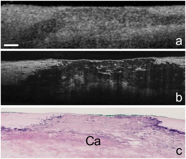
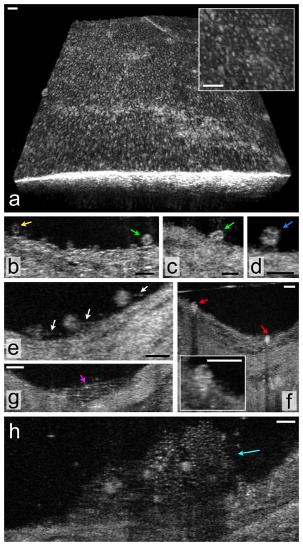
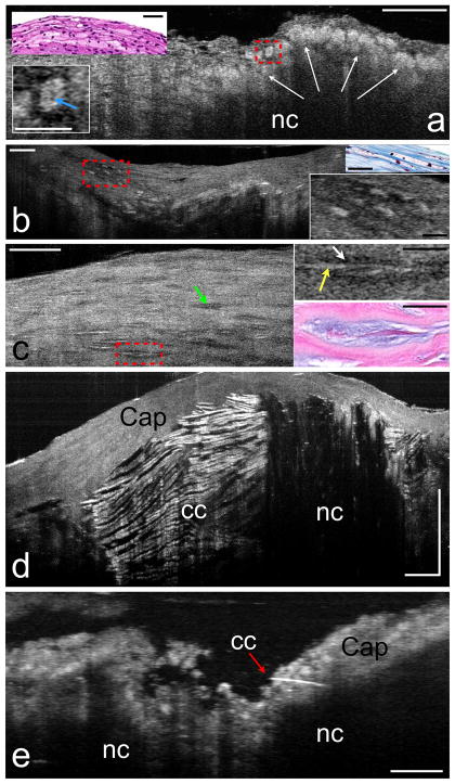
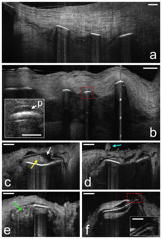
Progress in understanding, diagnosis, and treatment of coronary artery disease (CAD) has been hindered by our inability to observe cells and extracellular components associated with human coronary atherosclerosis in situ. The current standards for microstructural investigation, histology and electron microscopy are destructive and prone to artifacts. The highest-resolution intracoronary imaging modality, optical coherence tomography (OCT), has a resolution of ~10 μm, which is too coarse for visualizing most cells. Here we report a new form of OCT, termed micro-optical coherence tomography (μOCT), whose resolution is improved by an order of magnitude. We show that μOCT images of cadaver coronary arteries provide clear pictures of cellular and subcellular features associated with atherogenesis, thrombosis and responses to interventional therapy. These results suggest that μOCT can complement existing diagnostic techniques for investigating atherosclerotic specimens, and that μOCT may eventually become a useful tool for cellular and subcellular characterization of the human coronary wall in vivo.


Figures

Figure 1
Figure 1. μOCT images of a human coronary plaque
Human cadaver specimen. Comparison between corresponding OCT (a), μOCT (b), and histology images (c, Hematoxylin and Eosin) of a calcium plate (Ca) within the coronary artery wall. Scale bar, 200 μm.
Figure 2
Figure 2. μOCT of superficial arterial morphology
(a) Three-dimensional rendering of the swine coronary artery ex vivo, demonstrating a pattern of raised cells that are consistent with endothelial “pavementing”. (bh) Human cadaver specimens. (b) Multiple cells that are likely leukocytes (arrows) are seen adhering to the luminal surface in this μOCT image of a coronary plaque. Two different cell morphologies can be observed, one smaller cell with scant cytoplasm, consistent with a lymphocyte (yellow arrow) and another, slightly larger cell with a highly scattering, abundant cytoplasm, suggestive of a monocyte (green arrow). (c) Cell with an indented, bean-shaped nucleus (green arrow) characteristic of a monocyte. (d) Cell with a multi-lobed nucleus, possibly a neutrophil (blue arrow), is attached to the endothelial surface. (e) Multiple leukocytes tethered to the endothelial surface by linear structures suggestive of pseudopodia (white arrows). (f) Cells with the morphology of monocytes (red arrows) are seen in this cross-section and inset to be transmigrating through the endothelium. (g) Structures consistent with fibrin (magenta arrow) are visible as linear strands bridging a gap in the coronary artery wall. (h) Thrombus (cyan arrow) that appears to contain fibrin, small (2–3 μm diameter) highly scattering structures likely to be platelets, and multiple, entrapped cells. Scale bars, 30 μm.
Figure 3
Figure 3. μOCT of plaque morphology
Human cadaver specimens. (a) Necrotic core (nc) fibroatheroma with highly scattering lipid-laden macrophages or foam cells (white arrows) infiltrating the cap, also seen in the corresponding histology (upper left inset). An intracellular region of low μOCT signal, which may represent the nucleus, can be observed within the cytoplasm of some foam cells (e.g. lower left inset, blue arrow). (b) Another lesion, visualized by μOCT and histology, contains highly scattering foam cells that are ellipsoidal (right insets). (c) Smooth muscle cells by μOCT appear as spindle-shaped cells (green arrow). Smooth muscle cells producing collagen have a high backscattering interior (right upper inset, yellow arrow) and a “halo” of low backscattering (right upper inset, white arrow). Matching histology (right lower inset) demonstrates that the high backscattering region represents the cell body, while the lower intensity halo corresponds to collagen matrix. (d) Large necrotic core (nc) fibroatheroma, demonstrating cholesterol crystals (cc), characterized by reflections from their top and bottom surfaces. (e) A thin crystal (red arrow) appears to be piercing the cap of another necrotic core (nc) plaque. Scale bars for all primary images, 100 μm. Scale bars for all insets, 30 μm.
Figure 4
Figure 4. μOCT of stent and neointimal morphology
Human cadaver specimens. (a) μOCT image from a coronary segment with an implanted BMS shows struts devoid of polymer, covered by neointima. (b) DES struts from another cadaver showing polymer (red dashed box and p, inset) overlying the strut reflections. (c) Tissue (yellow arrow) is interposed between the polymer and the stent strut and the polymer has fractured (white arrow). (d) Superficial leukocyte cluster (cyan arrow) and adjacent attached leukocytes overlying the site of the polymer fracture. (e) Apparent inflammation at the edge of a strut (green arrow). (f) Uncovered strut, completely devoid of overlying endothelium (red dashed box and inset). Scale bars for primary images, 100 μm. Scale bars for all insets, 30 μm.

References

    1. Virmani R, Kolodgie FD, Burke AP, Farb A, Schwartz SM. Lessons from sudden coronary death - A comprehensive morphological classification scheme for atherosclerotic lesions. Arteriosclerosis Thrombosis and Vascular Biology. 2000;20:1262–1275. - PubMed
    1. Stary HC, et al. A Definition of Advanced Types of Atherosclerotic Lesions and a Histological Classification of Atherosclerosis: A Report From the Committee on Vascular Lesions of the Council on Arteriosclerosis, American Heart Association. Circulation. 1995;92:1355–1374. - PubMed
    1. Gronholdt MLM, Dalager-Pedersen S, Falk E. Coronary atherosclerosis: determinants of plaque rupture. European Heart Journal. 1998;19:C24–C29. - PubMed
    1. Davies MJ. Acute coronary thrombosis - The role of plaque disruption and its initiation and prevention. European Heart Journal. 1995;16:3–7. - PubMed
    1. Pasternak RC, Baughman KL, Fallon JT, Block PC. Scanning electron microscopy after coronary transluminal angioplasty of normal canine coronary arteries. American Journal of Cardiology. 1980;45 - PubMed

Publication types