. 2011 Jul 10;8(8):691-6.
doi: 10.1038/nmeth.1649.

Protein standard absolute quantification (PSAQ) method for the measurement of cellular ubiquitin pools

Affiliations

Protein standard absolute quantification (PSAQ) method for the measurement of cellular ubiquitin pools

Stephen E Kaiser et al. Nat Methods. .

Abstract

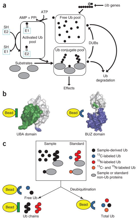
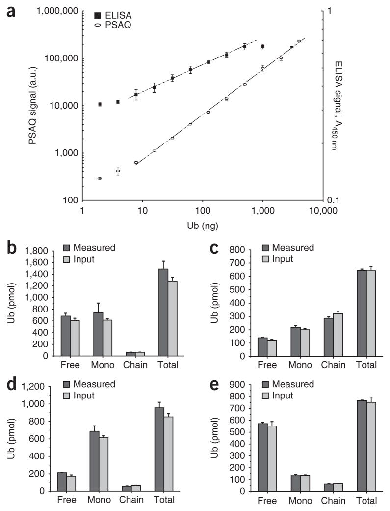
The protein ubiquitin is an important post-translational modifier that regulates a wide variety of biological processes. In cells, ubiquitin is apportioned among distinct pools, which include a variety of free and conjugated species. Although maintenance of a dynamic and complex equilibrium among ubiquitin pools is crucial for cell survival, the tools necessary to quantify each cellular ubiquitin pool have been limited. We have developed a quantitative mass spectrometry approach to measure cellular concentrations of ubiquitin species using isotope-labeled protein standards and applied it to characterize ubiquitin pools in cells and tissues. Our method is convenient, adaptable and should be a valuable tool to facilitate our understanding of this important signaling molecule.


Conflict of interest statement

COMPETING FINANCIAL INTERESTS

The authors declare no competing financial interests.

Figures

Figure 1
Figure 1
Ubiquitin pools and assay overview. (a) Ubiquitin (Ub) is present in eukaryotic cells as a mixture of free ubiquitin, monoubiquitinated substrates, polyubiquitin chains and activated species linked to enzymes by thioester bonds. DUBs, deubiquitinating enzymes. E1, ubiquitin activating enzyme. E2, ubiquitin transfer enzyme. SH, catalytic cysteine residues; S, catalytic cysteine residues conjugated via covalent thioester linkages to ubiquitin. (b) Cartoon representations of the interactions of the protein-affinity reagents used in the Ub-PSAQ assay with ubiquitin. The hP2 UBA domain binds the Ile44-centered hydrophobic patch on ubiquitin (patch is obscured on the left and is shown in green on the right) and preferentially binds ubiquitin chains without bias for specific ubiquitin-ubiquitin linkages,,. The BUZ domain of IsoT binds the C-terminal Gly-Gly of free ubiquitin species (shown in blue on the left). (c) Schematic of the Ub-PSAQ assay. Isotope-labeled protein standards are added to lysates containing sample derived ubiquitin species. Half of the sample is treated with the deubiquitinating enzyme usp2cc, free ubiquitin species are bound to the BUZ affinity reagent, washed, eluted and then trypsinized before quantification of total sample–derived ubiquitin relative to the 13C-labeled free-ubiquitin protein standard by LC-ESI TOF MS. Free ubiquitin species in the untreated half of the sample are similarly captured by the BUZ affinity reagent and quantified by LC-ESI MS. Next, the untreated half of the sample is incubated with the hP2 UBA affinity reagent to capture polyubiquitin chains, which are quantified relative to the polyubiquitin chain standard.
Figure 2
Figure 2
Assay validation. (a) Superimposed serial dilution curves of ubiquitin detection by BUZ-enriched PSAQ and ELISA comparing the linear dynamic range and the lower limit of detection. Mass spectrometry signal intensity is expressed in arbitrary units (a.u.) after normalization to the spiked 13C-labeled free-ubiquitin standard. A450 nm, absorbance at 450 nm. Data are represented as mean ± s.d. (n = 8 for ELISA and n = 3 for PSAQ). (be) Measurement of defined mixtures composed of unlabeled free ubiquitin, autoubiquitinated Rsp5 and ubiquitin-GFP mock monoubiquitinated substrate containing similar amounts of input free ubiquitin and ubiquitin-GFP substrate (b), higher levels of ubiquitin-GFP and ubiquitin chains (chain) than free ubiquitin (c), high levels of mock monoubiquitinated substrate (mono) (d) and higher levels of free ubiquitin (e). Error bars (be), ± s.d. (n = 3).
Figure 3
Figure 3
Effect of acute proteasome inhibition on ubiquitin pools. (a,b) Ub-PSAQ analysis of lysates from HEK293 (a) and MEF (b) cell lines treated with either DMSO (vehicle) or the proteasome inhibitor MG-132 (1 μM) over 12 h. Error bars, ± s.d. (n = 3). *P < 0.05; **P < 0.01; and ***P < 0.005 (unpaired t-test). MonoUb, monoubiquitin conjugates. (c) Representation of ubiquitin pool components in HEK293 and MEF cell lines. (d) Ubiquitin western blots with antibodies FK2 and A100 showing ubiquitin-immunoreactive material for HEK293 cells treated with either DMSO (vehicle) or MG-132 (1 μM) over 12 h. Bars on top right of blots denote high-molecular-weight ubiquitin conjugates (HMW Ub conjugates), and ubiquitin-modified histone H2A (UH) and free ubiquitin are indicated. (e) Distribution of monoubiquitinated substrates in cytosolic and histone-enriched fractions from HEK293 cells treated with either DMSO or MG-132 (10 μM) for 6 h.
Figure 4
Figure 4
Ub-PSAQ analysis of ubiquitin pools in cytosolic and histone-enriched fractions from HEK293 cells. (a) Representation of ubiquitin pool components in cytosolic and histone-enriched fractions. (b) Distribution of ubiquitin pool components in cytosolic and histone-enriched fractions. Bar graphs show concentration of the indicated species per milligram of total protein in each fraction. Pie charts show the same data after normalization to show the distribution of each species. (c) Distribution of ubiquitin chain linkages in cytosolic and histone-enriched fractions. Bar graphs show the concentration of each species within each fraction, and pie charts show the distribution of each species between cytosolic and histone-enriched fractions. Error bars (b,c), means ± s.d. (n = 3).

Comment in

References

    1. Ravid T, Hochstrasser M. Diversity of degradation signals in the ubiquitin-proteasome system. Nat Rev Mol Cell Biol. 2008;9:679–690. - PMC - PubMed
    1. Mukhopadhyay D, Riezman H. Proteasome-independent functions of ubiquitin in endocytosis and signaling. Science. 2007;315:201–205. - PubMed
    1. Zeng W, et al. Reconstitution of the RIG-I pathway reveals a signaling role of unanchored polyubiquitin chains in innate immunity. Cell. 2010;141:315–330. - PMC - PubMed
    1. Hoeller D, Dikic I. Targeting the ubiquitin system in cancer therapy. Nature. 2009;458:438–444. - PubMed
    1. Lee BH, et al. Enhancement of proteasome activity by a small-molecule inhibitor of USP14. Nature. 2010;467:179–184. - PMC - PubMed

Publication types