. 2011 Jul 10;475(7356):353-8.
doi: 10.1038/nature10238.

The crystal structure of a voltage-gated sodium channel

Affiliations

The crystal structure of a voltage-gated sodium channel

Jian Payandeh et al. Nature. .

Abstract

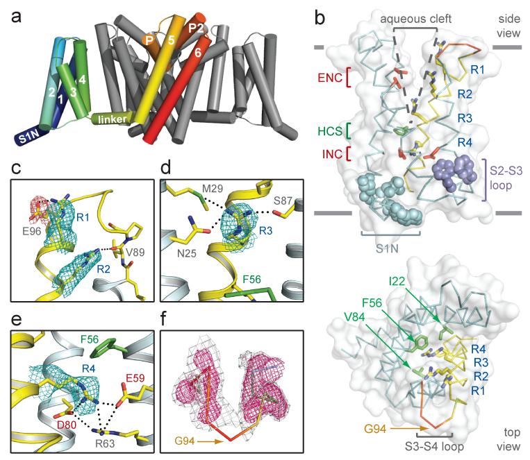
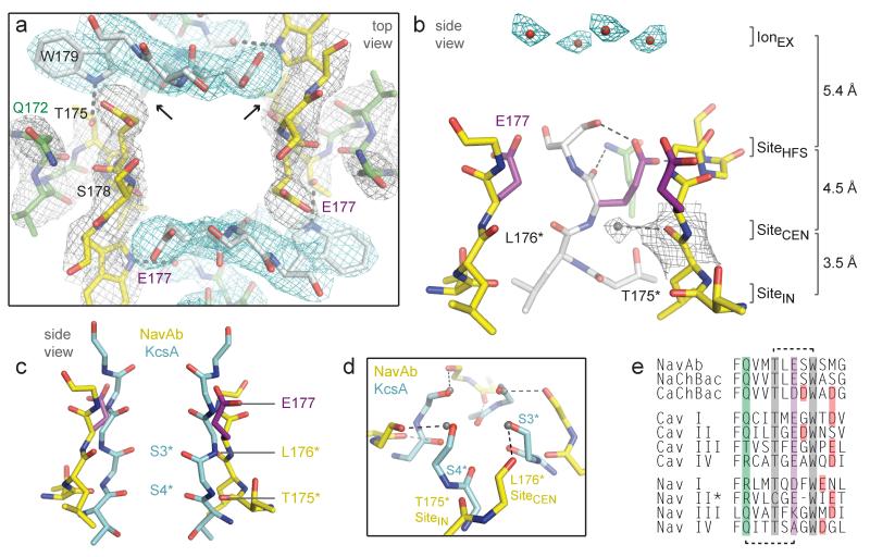
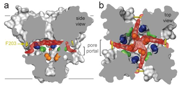
Voltage-gated sodium (Na(V)) channels initiate electrical signalling in excitable cells and are the molecular targets for drugs and disease mutations, but the structural basis for their voltage-dependent activation, ion selectivity and drug block is unknown. Here we report the crystal structure of a voltage-gated Na(+) channel from Arcobacter butzleri (NavAb) captured in a closed-pore conformation with four activated voltage sensors at 2.7 Å resolution. The arginine gating charges make multiple hydrophilic interactions within the voltage sensor, including unanticipated hydrogen bonds to the protein backbone. Comparisons to previous open-pore potassium channel structures indicate that the voltage-sensor domains and the S4-S5 linkers dilate the central pore by pivoting together around a hinge at the base of the pore module. The NavAb selectivity filter is short, ∼4.6 Å wide, and water filled, with four acidic side chains surrounding the narrowest part of the ion conduction pathway. This unique structure presents a high-field-strength anionic coordination site, which confers Na(+) selectivity through partial dehydration via direct interaction with glutamate side chains. Fenestrations in the sides of the pore module are unexpectedly penetrated by fatty acyl chains that extend into the central cavity, and these portals are large enough for the entry of small, hydrophobic pore-blocking drugs. This structure provides the template for understanding electrical signalling in excitable cells and the actions of drugs used for pain, epilepsy and cardiac arrhythmia at the atomic level.


Figures

Figure 1
Figure 1. Structure of NavAb and the activated VSD
a, Structural elements in NavAb. One subunit is highlighted (1-6, transmembrane segments S1-S6). The nearest VSD has been removed for clarity. b, Side and top-views of the VSD illustrating the extracellular negative charge-cluster (red, ENC), the intracellular negative charge-cluster (red, INC), hydrophobic constriction site (green, HCS), residues of the S1N helix (cyan) and phenylalanines of the S2-S3 loop (purple). S4 segment and gating charges (R1-R4) are in yellow. c-e, Hydrogen bonding of gating-charges, dotted lines (<3.5 Å). Fo-Fc omit maps are contoured over E96 and R1-R4 at 1, 1, 1.5, 2.5 and 1.75 σ, respectively. f, S3-S4 loop. Colored according to crystallographic temperature factors of the main-chain (blue < 50 Å2 to red > 150 Å2). An Fo-Fc omit map is contoured at 1.5 σ (grey) and 2.5 σ (pink).
Figure 2
Figure 2. NavAb pore module
a, Pore-lining S6 helices of NavAb (yellow) and the closed MlotiK (3BEH), KcsA (1K4C) and NaK (2AHY) channels. Cα locations of Met221 defines a common radius for the closed activation gate (red circle). b, Comparison of S6 helices of NavAb and KV1.2/2.1 (2R9R). c, Interaction of S6 with S4-S5 linkers (NavAb, top; KV1.2/2.1, bottom). d, Architecture of the NavAb pore. Glu177 side-chains (purple sticks); pore volume, grey. e, Electrostatic potential colored from −10 to 10 kT (red to blue).
Figure 3
Figure 3. Structure of the NavAb selectivity filter
a, Top view of the selectivity filter. Symmetry-related molecules are colored white and yellow; P-helix residues are colored green. Hydrogen bonds between Thr175 and Trp179 are indicated by grey dashes. Electron-densities from Fo-Fc omit maps are contoured at 4.0 σ (blue and grey) and subtle differences can be appreciated (small arrows). b, Side view of the selectivity filter. Glu177 (purple) interactions with Gln172, Ser178 and the backbone of Ser180 are shown in the far subunit. Fo-Fc omit map, 4.75 σ (blue); putative cations or water molecules (red spheres, IonEX). Electron-density around Leu176 (grey; Fo-Fc omit map at 1.75 σ) and a putative water molecule is shown (grey sphere). Na+-coordination sites: SiteHFS, SiteCEN and SiteIN. c, Superposition of NavAb and a K+-channel selectivity filter: NavAb Glu177 side-chains (purple) and backbone carbonyls (*); K+-channel (blue, 1K4C), site 3 and site 4 backbone carbonyls (ref. 35) (*). This structural alignment is based on P-helices. d, Enlarged view of SiteCEN and SiteIN: water molecules, grey spheres; dotted lines, ~2.5 Å. e, Selectivity filter sequence alignment. E177 homologs, purple; outer-ring of negatively charged residues (red).
Figure 4
Figure 4. Membrane access to the central cavity in NavAb
a, Side-view through the pore module illustrating fenestrations (portals) and hydrophobic access to central cavity. Phe203 side-chains, yellow sticks. Surface representations of NavAb residues aligning with those implicated in drug binding and block, Thr206, blue; Met209, green; Val213, orange. Membrane boundaries, grey lines. Electron-density from an Fo-Fc omit map is contoured at 2.0 σ. b, Top-view sectioned below the selectivity filter, colored as in a.
Figure 5
Figure 5. Model for activation gate opening
a, Superposition of the NavAb and KV1.2/2.1 based on their VSDs (cylinders). b, Superposition of NavAb and KV1.2/2.1 tetrameric pore modules (PM) viewed from the membrane: S5 gating hinge, *. c-d, S1 interaction with P-helix. .S1 Thr is P-helix is 2.9 Å neighboring subunit in KV1.2/2.1 (), but >4.5 Å in NavAb.

Comment in

Similar articles

Cited by

References

    1. Koth CM, Payandeh J. Strategies for the cloning and expression of membrane proteins. Adv Protein Chem Struct Biol. 2009;76:43–86. - PubMed
    1. Cronin CN, Lim KB, Rogers J. Production of selenomethionyl-derivatized proteins in baculovirus-infected insect cells. Protein Sci. 2007;16:2023–2029. - PMC - PubMed
    1. Chaptal V, et al. Fluorescence Detection of Heavy Atom Labeling (FD-HAL): A rapid method for identifying covalently modified cysteine residues by phasing atoms. J Struct Biol. 2010;171:82–87. - PubMed
    1. Faham S, Bowie JU. Bicelle crystallization: a new method for crystallizing membrane proteins yields a monomeric bacteriorhodopsin structure. J Mol Biol. 2002;316:1–6. - PubMed
    1. Faham S, et al. Crystallization of bacteriorhodopsin from bicelle formulations at room temperature. Protein Sci. 2005;14:836–840. - PMC - PubMed

Publication types

MeSH terms