

Lentiviral Based Gene Transduction and Promoter Studies in Human Hematopoietic Stem Cells (hHSCs)
- PMID: 21743782
- PMCID: PMC3130352
- DOI: 10.46582/jsrm.0701005
Lentiviral Based Gene Transduction and Promoter Studies in Human Hematopoietic Stem Cells (hHSCs)
Abstract
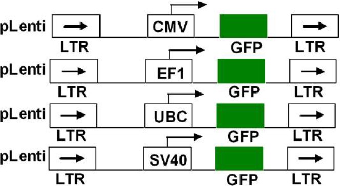
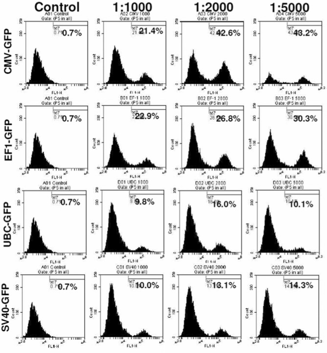
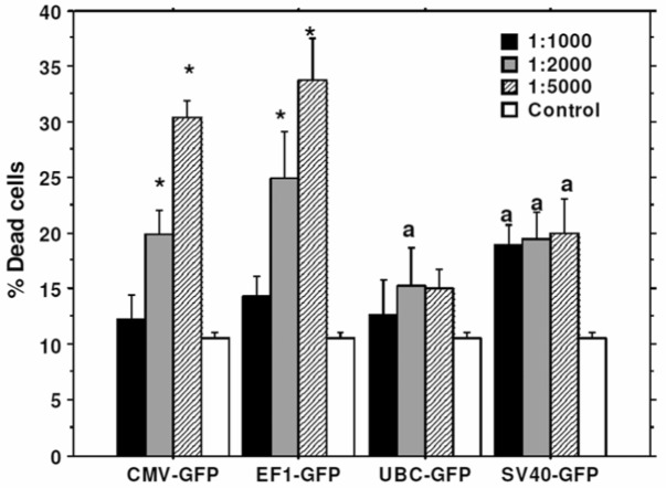
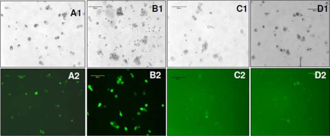
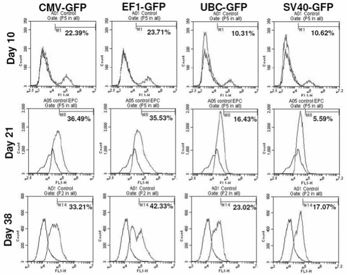
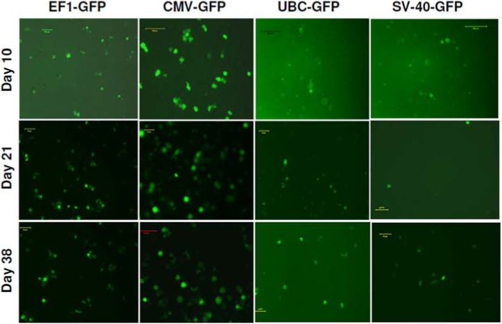
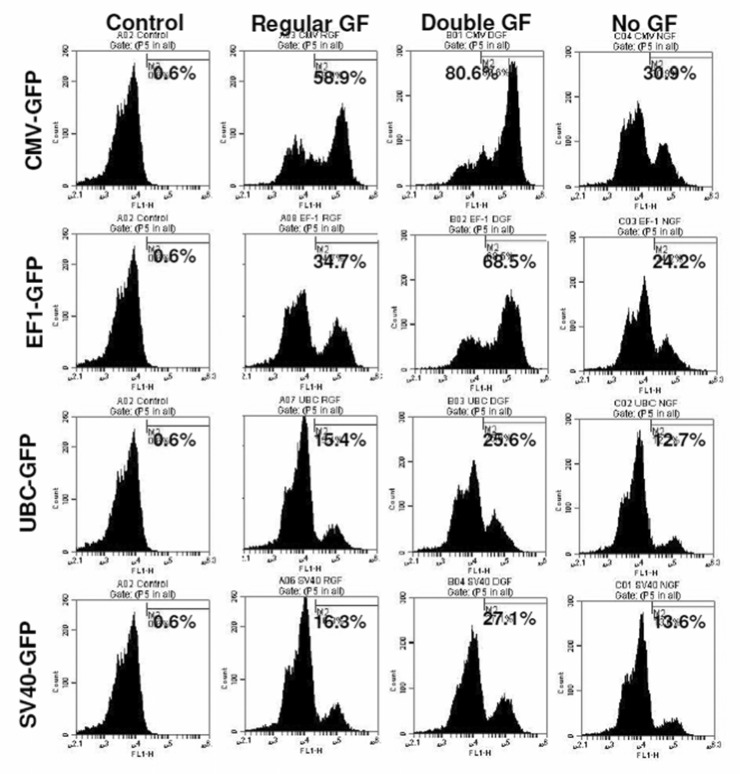
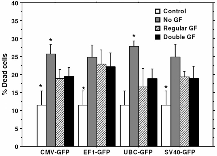
Human hematopoietic stem cells (hHSCs) have enormous potential for clinical use in cell-based therapies, especially as a gene delivery system. Moreover, lentiviral transduction in stem cells is very often associated with low transduction efficiency and low levels of foreign gene expression. Therefore, it is important to analyze vector and promoter systems that can generate robust foreign gene expression in these cells. In this study, we evaluated and compared the ability of different commercially available promoters to drive the expression of exogenous reporter genes in hHSCs and evaluated the effect of different doses of stem cell growth factors on the expression of transgenes. We used lentivirus based vector system carrying the following promoters: 1) Human cytomegalovirus (CMV) promoter, 2) Simian virus 40 (SV40) promoter, 3) mammalian Ubiquitin C (UBC) promoter and 4) cellular polypeptide chain elongation factor 1 alpha (EF1) promoter. EF1 and CMV promoters robustly drove the expression of green fluorescence protein (GFP) reporter gene, while SV40 and UBC promoters induced very low level of GFP expression. Lentivectors containing EF1 and CMV promoters showed high-level stable GFP expression in human cord blood stem cells for 6 weeks period after post transduction. CD133+ hHSCs stimulated with higher concentration of growth factors exhibited enhancement of transduction rate. Cord blood derived CD133+ hHSCs could be effectively transduced with lentivectors under CMV or EF-1 promoters for the expression of foreign gene.
Keywords: Cord blood stem cells (AC133+); Lentiviral vectors; Promoters and Green fluorescent protein (GFP).
Figures
References
-
- Fibbe WE, Noort WA, Schipper F, Willemze R: Ex vivo expansion and engraftment potential of cord blood-derived CD34+ cells in NOD/SCID mice. Ann N Y Acad Sci 2001, 938:9-17 - PubMed
-
- Hao QL, Shah AJ, Thiemann FT, Smogorzewska EM, Crooks GM: A functional comparison of CD34 + CD38- cells in cord blood and bone marrow. Blood 1995, 86:3745-3753 - PubMed
-
- Aker M, Varadi G, Slavin S, Nagler A: Fludarabine-based protocol for human umbilical cord blood transplantation in children with Fanconi anemia. J Pediatr Hematol Oncol 1999, 21:237-239 - PubMed
-
- Bielorai B, Hughes MR, Auerbach AD, Nagler A, Loewenthal R, Rechavi G, Toren A: Successful umbilical cord blood transplantation for Fanconi anemia using preimplantation genetic diagnosis for HLA-matched donor. Am J Hematol 2004, 77:397-399 - PubMed
-
- Kohn DB: Gene therapy for haematopoietic and lymphoid disorders. Clin Exp Immunol 1997, 107Suppl 1:54-57 - PubMed
Grants and funding
LinkOut - more resources
Full Text Sources
Other Literature Sources
Research Materials