. 2011;2(1):34-49.

TRANSCRIPTIONAL AND PHOSPHO-PROTEOMIC SCREENS REVEAL STEM CELL ACTIVATION OF INSULIN-RESISTANCE AND TRANSFORMATION PATHWAYS FOLLOWING A SINGLE MINIMALLY TOXIC EPISODE OF ROS

Affiliations

TRANSCRIPTIONAL AND PHOSPHO-PROTEOMIC SCREENS REVEAL STEM CELL ACTIVATION OF INSULIN-RESISTANCE AND TRANSFORMATION PATHWAYS FOLLOWING A SINGLE MINIMALLY TOXIC EPISODE OF ROS

R Mouzannar et al. Int J Genomics Proteomics. 2011.

Abstract

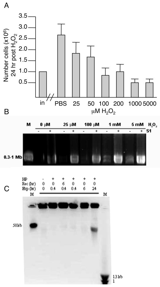
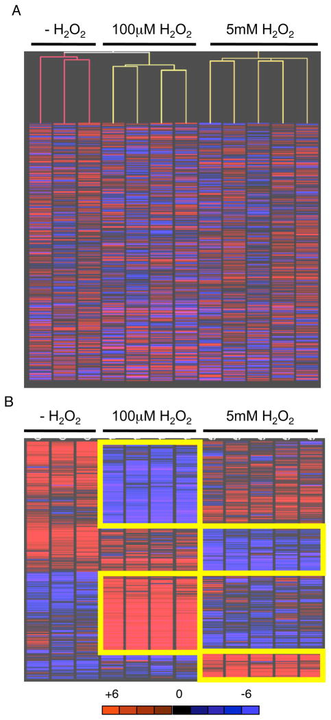
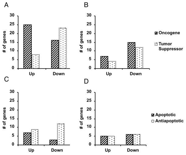
Elevated reactive oxidative species (ROS) are cytotoxic, and chronic elevated levels of ROS have been implicated in multiple diseases as well as cellular transformation and tumor progression. However, the potential for a transient and minimally toxic episode of ROS exposure, or a minimal threshold dose of ROS, to initiate disease or cellular transformation is unclear. We examined both transcriptional and phospho-proteomic responses of murine embryonic stem (ES) cells to a single brief exposure of minimally toxic hydrogen peroxide (H(2)O(2)). The cellular response was distinct from those induced by either an acute exposure to H(2)O(2) or the topoisomerase II poison etoposide. Analysis of tumorigenesis-related transcripts revealed a significant up-regulation of oncogenes and down-regulation of tumor suppressors. Analysis of the phospho-proteomic response demonstrated insulin-signaling induction, including insulin receptor Y972 hypophosphorylation, similar to insulin-resistance mouse models and observed in diabetic patients. In addition, ES cells were more resistant to ROS than differentiated cells, and retained their transcriptional self-renewal signature, suggesting stem cells have a higher potential for ROS-mediated mutagenesis and proliferation in vivo. These results are a direct demonstration that even brief and non-toxic exposures to ROS may induce transduction of insulin resistance and transformation signaling in stem cells leading to diabetes and cancer.


Figures

Fig. 1
Fig. 1
Response of ES cells in culture 24 hrs following exposure to minimally toxic ROS. A. Cells were exposed to H2O2 for 15 min, recovered in normal medium for 20 hrs, then the number of viable cells in culture scored (dead cells were excluded by trypan blue). Data are the average and standard deviation of at least 4 independent experiments. B. Dose-dependent accumulation of single-strand breaks (SSBs) following exposure to H2O2 for 15 min. Pulse field gel electrophoresis of agarose embedded DNA without (−; DSB) or with (+; SSB and DSB) S1 nuclease digestion. DNA fragmentation was not evident in samples exposed to H2O2 at lower doses. Fragmentation was observed at higher doses (1mM and 5mM). As expected, visible fragmentation was detected in all samples, including untreated, following S1 nuclease digestion. Visible fragments were within the 0.3-1 Mb range. C. Time-dependent accumulation of 50 kb fragments following exposure (Exp) to 100 M H2O2 with or without recovery (Rec). 50 kb fragments observed only after chronic 24 hour continuous exposure and no recovery. M -- size standard marker.
Fig. 2
Fig. 2
A. Relatedness of sample groups confirmed by unsupervised Pearson correlation clustering of transcripts deregulated by two-fold between controls and replicate samples. Analysis performed by GeneSpring data analysis software. B. Distinct gene expression signatures induced by low and high doses of H2O2. Parametric t-test with unequal variance (Welch t-test) analysis was performed by GeneSpring data analysis software.
Fig. 3
Fig. 3
IPA clustering shows distinct response of ES cells to minimally toxic ROS. IPA clustering was performed on statistically significant altered transcripts from four samples exposed to 100uM H2O2, five samples exposed to 5mM H2O2, and three replicate control samples analyzed by Affymetrix MG-U74VerA chips and GeneSpring software. A. Altered oncogene and tumor suppressor transcripts following 100 M H2O2. B. Altered oncogene and tumor suppressor transcripts following 5mM H2O2. C. Altered apoptotic and anti-apoptotic transcripts following 100 M H2O2. D. Altered apoptotic and anti-apoptotic transcripts following 5mM H2O2.

Similar articles

Cited by

References

    1. Jackson AL, Loeb LA. Mutation Res. 2001;477:7–21. - PubMed
    1. Davies KJ. Biochem Soc Symp. 1995;61:1–31. - PubMed
    1. Beckman KB, Ames BN. J Biol Chem. 1997;272:19633–6. - PubMed
    1. Waris G, Ahsan H. J Carcinog. 2006;5:14. - PMC - PubMed
    1. Burdon RH. Free Radio Biol Med. 1995;18:775–94. - PubMed